| Description | Sets of proteins participating in pathways from KEGG |
| Measurement | association by literature curation |
| Association | protein-pathway associations from curated pathways |
| Category | structural or functional annotations |
| Resource | Kyoto Encyclopedia of Genes and Genomes |
| Citation(s) |
|
| Last Updated | Thu Jan 08 00:00:00 UTC 2026 |
| Stats |
|
| API | |
| Script | |
| Downloads |
Gene Attribute

Gene Similarity
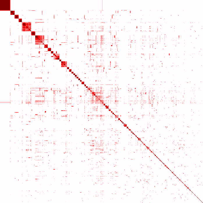
Attribute Similarity
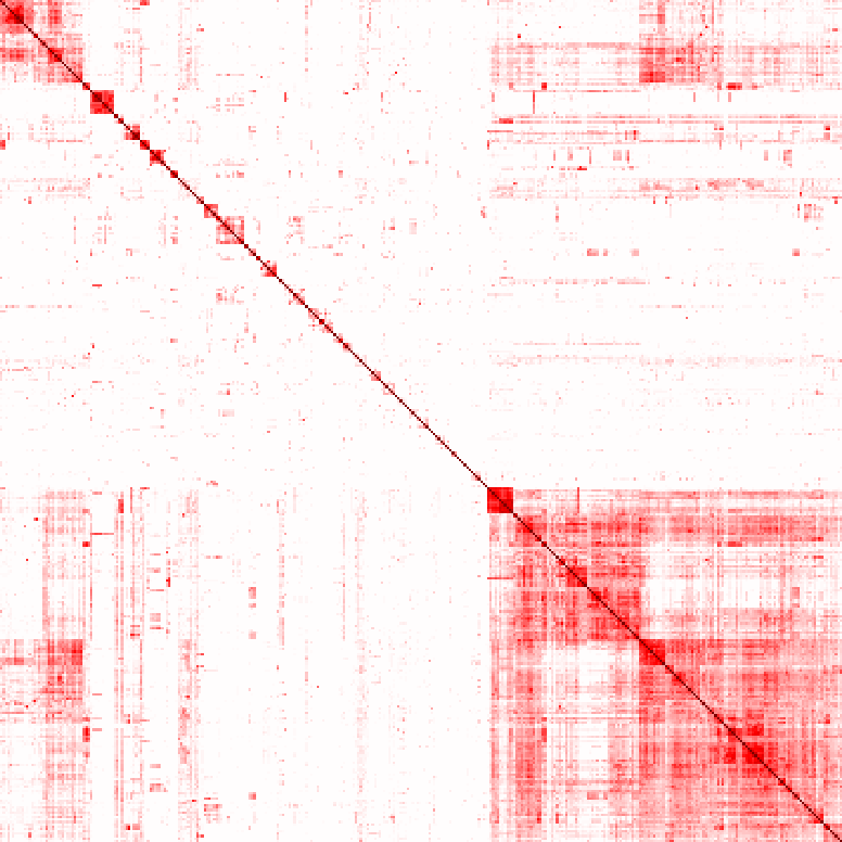
UMAP
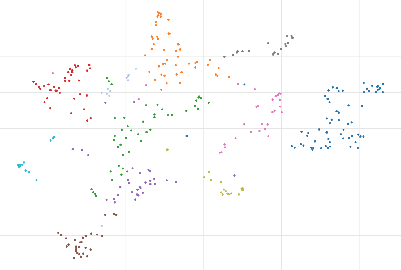
356 sets of proteins participating in pathways from the KEGG Pathways 2026 dataset.
| Gene Set | Description |
|---|---|
| Glycolysis / Gluconeogenesis | Glycolysis is the process of converting glucose into pyruvate and generating small amounts of ATP (energy) and NADH (reducing power). It is a central pathway that produces important precursor metabolites: six-carbon compounds of glucose-6P and fructose-6P and three-carbon compounds of glycerone-P, glyceraldehyde-3P, glycerate-3P, phosphoenolpyruvate, and pyruvate [MD:M00001]. Acetyl-CoA, another important precursor metabolite, is produced by oxidative decarboxylation of pyruvate [MD:M00307]. When the enzyme genes of this pathway are examined in completely sequenced genomes, the reaction steps of three-carbon compounds from glycerone-P to pyruvate form a conserved core module [MD:M00002], which is found in almost all organisms and which sometimes contains operon structures in bacterial genomes. Gluconeogenesis is a synthesis pathway of glucose from noncarbohydrate precursors. It is essentially a reversal of glycolysis with minor variations of alternative paths [MD:M00003]. |
| Citrate cycle (TCA cycle) | The citrate cycle (TCA cycle, Krebs cycle) is an important aerobic pathway for the final steps of the oxidation of carbohydrates and fatty acids. The cycle starts with acetyl-CoA, the activated form of acetate, derived from glycolysis and pyruvate oxidation for carbohydrates and from beta oxidation of fatty acids. The two-carbon acetyl group in acetyl-CoA is transferred to the four-carbon compound of oxaloacetate to form the six-carbon compound of citrate. In a series of reactions two carbons in citrate are oxidized to CO2 and the reaction pathway supplies NADH for use in the oxidative phosphorylation and other metabolic processes. The pathway also supplies important precursor metabolites including 2-oxoglutarate. At the end of the cycle the remaining four-carbon part is transformed back to oxaloacetate. According to the genome sequence data, many organisms seem to lack genes for the full cycle [MD:M00009], but contain genes for specific segments [MD:M00010 M00011]. |
| Pentose phosphate pathway | |
| Pentose and glucuronate interconversions | |
| Fructose and mannose metabolism | |
| Galactose metabolism | |
| Ascorbate and aldarate metabolism | |
| Starch and sucrose metabolism | |
| Pyruvate metabolism | |
| Glyoxylate and dicarboxylate metabolism | |
| Propanoate metabolism | |
| Butanoate metabolism | |
| Inositol phosphate metabolism | |
| Oxidative phosphorylation | The phosphorylation of ADP to ATP that accompanies the oxidation of a metabolite through the operation of the respiratory chain. Oxidation of compounds establishes a proton gradient across the membrane, providing the energy for ATP synthesis. |
| Nitrogen metabolism | |
| Sulfur metabolism | Sulfur is an essential element for life and the metabolism of organic sulfur compounds plays an important role in the global sulfur cycle. Sulfur occurs in various oxidation states ranging from +6 in sulfate to -2 in sulfide (H2S). Sulfate reduction can occur in both an energy consuming assimilatory pathway and an energy producing dissimilatory pathway. The assimilatory pathway, which is found in a wide range of organisms, produces reduced sulfur compounds for the biosynthesis of S-containing amino acids and does not lead to direct excretion of sulfide. In the dissimilatory pathway, which is restricted to obligatory anaerobic bacterial and archaeal lineages, sulfate (or sulfur) is the terminal electron acceptor of the respiratory chain producing large quantities of inorganic sulfide. Both pathways start from the activation of sulfate by reaction with ATP to form adenylyl sulfate (APS). In the assimilatory pathway [MD:M00176] APS is converted to 3'-phosphoadenylyl sulfate (PAPS) and then reduced to sulfite, and sulfite is further reduced to sulfide by the assimilatory sulfite reductase. In the dissimilatory pathway [MD:M00596] APS is directly reduced to sulfite, and sulfite is further reduced to sulfide by the dissimilatory sulfite reductase. The capacity for oxidation of sulfur is quite widespread among bacteria and archaea, comprising phototrophs and chemolithoautotrophs. The SOX (sulfur-oxidation) system [MD:M00595] is a well-known sulfur oxidation pathway and is found in both photosynthetic and non-photosynthetic sulfur-oxidizing bacteria. Green sulfur bacteria and purple sulfur bacteria carry out anoxygenic photosynthesis with reduced sulfur compounds such as sulfide and elemental sulfur, as well as thiosulfate (in some species with the SOX system), as the electron donor for photoautotrophic growth. In some chemolithoautotrophic sulfur oxidizers (such as Thiobacillus denitrificans), it has been suggested that dissimilatory sulfur reduction enzymes operate in the reverse direction, forming a sulfur oxidation pathway from sulfite to APS and then to sulfate. |
| Fatty acid biosynthesis | |
| Fatty acid elongation | The elongation of a fatty acid chain by the sequential addition of two-carbon units. |
| Fatty acid degradation | |
| Steroid biosynthesis | |
| Primary bile acid biosynthesis | Bile acids are steroid carboxylic acids derived from cholesterol in vertebrates. The primary bile acids, cholic acid and chenodeoxycholic acid, are synthesized in the liver and conjugated with taurine or glycine before secretion via bile into the intestine. The conversion from cholesterol to cholic and chenodeoxycholic acids involves four steps: 1) the initiation of synthesis by 7alpha-hydroxylation of sterol precursors, 2) further modifications to the ring structures, 3) side-chain oxidation and shortening (cleavage) by three carbons, and 4) conjugation of the bile acid with taurine or glycine. |
| Steroid hormone biosynthesis | Steroid hormones derived from cholesterol are a class of biologically active compounds in vertebrates. The cholesterol side-chain cleavage enzyme CYP11A1 catalyzes conversion of cholesterol, a C27 compound, to the first C21 steroid, pregnenolone, which is converted by a bifunctional enzyme complex to the gestagen hormone, progesterone [MD:M00107]. Pregnenolone and progesterone are the starting materials for the three groups of steroids: C21 steroids of glucocorticoids and mineralocorticoids, C19 steroids of androgens, and C18 steroids of estrogens. (i) Progesterone is converted by hydroxylations at carbons 21 and 11 to corticosterone, which is further modified by hydroxylation and oxydoreduction at carbon 18 to yield aldosterone, a mineralcorticoid [MD:M00108]. Cortisol, the main glucocorticoid, is formed from 17alpha-hydroxyprogesterone with 11-deoxycortisol as an intermediate [MD:M00109]. (ii) Male hormone testosterone is formed from pregnenolone by two pathways, delta5 pathway via dehydroepiandrosterone and delta4 pathway via androstenedione [MD:M00110]. The enzyme CYP17A1 is responsible for the 17,20 lyase and 17alpha-hydroxylase activities in respective pathways. (iii) Female hormones estrone and estradiol are formed from testosterone and 4-androstene-3,17-dione by oxidative removal of the C19 methyl group and subsequent aromatization of ring A. In addition to these three groups, recent studies show that there is another group, termed neurosteroids, synthesized in the brain rather than the peripheral endocrine gland. |
| Glycerolipid metabolism | |
| Glycerophospholipid metabolism | |
| Ether lipid metabolism | |
| Sphingolipid metabolism | |
| Arachidonic acid metabolism | |
| Linoleic acid metabolism | |
| alpha-Linolenic acid metabolism | |
| Biosynthesis of unsaturated fatty acids | |
| Purine metabolism | |
| Pyrimidine metabolism | |
| Alanine, aspartate and glutamate metabolism | |
| Glycine, serine and threonine metabolism | Serine is derived from 3-phospho-D-glycerate, an intermediate of glycolysis [MD:M00020], and glycine is derived from serine. Threonine is an essential amino acid, which animals cannot synthesize. In bacteria and plants, threonine is derived from aspartate [MD:M00018]. |
| Cysteine and methionine metabolism | Cysteine and methionine are sulfur-containing amino acids. Cysteine is synthesized from serine through different pathways in different organism groups. In bacteria and plants, cysteine is converted from serine (via acetylserine) by transfer of hydrogen sulfide [MD:M00021]. In animals, methionine-derived homocysteine is used as sulfur source and its condensation product with serine (cystathionine) is converted to cysteine [MD:M00338]. Cysteine is metabolized to pyruvate in multiple routes. Methionine is an essential amino acid, which animals cannot synthesize. In bacteria and plants, methionine is synthesized from aspartate [MD:M00017]. S-Adenosylmethionine (SAM), synthesized from methionine and ATP, is a methyl group donor in many important transfer reactions including DNA methylation for regulation of gene expression. SAM may also be used to regenerate methionine in the methionine salvage pathway [MD:M00034]. |
| Valine, leucine and isoleucine degradation | |
| Valine, leucine and isoleucine biosynthesis | |
| Lysine degradation | |
| Arginine biosynthesis | |
| Arginine and proline metabolism | |
| Histidine metabolism | |
| Tyrosine metabolism | |
| Phenylalanine metabolism | |
| Tryptophan metabolism | |
| Phenylalanine, tyrosine and tryptophan biosynthesis | |
| beta-Alanine metabolism | |
| Taurine and hypotaurine metabolism | |
| Phosphonate and phosphinate metabolism | Natural products containing carbon-phosphorous bonds, so-called C-P compounds, are derivatives of phosphonate and phosphinate with substitution of alkyl group for hydrogen of phosphorus-hydrogen bonds. C-P compounds have been found in many organisms, but only protists and bacteria, mostly Actinobacteria, have biosynthetic capacity. A common reaction in the biosynthetic pathway is C-P bond forming reaction from phosphoenolpyruvate (PEP) to phosphonopyruvate (PnPy) catalyzed by PEP phosphomutase. 2-Aminoethylphosphonate (AEP) is the most abundant C-P compound in the natural world. AEP derivatives include phosphonoprotein, phosphonoglycan, and phosphonolipid. Other known C-P compounds are bioactive substances used in medicine (antibiotics) and agriculture (herbicide) such as fosfomycin, FR-33289, rhizocticin, and bialaphos. |
| Selenocompound metabolism | |
| D-Amino acid metabolism | |
| Glutathione metabolism | |
| Amino sugar and nucleotide sugar metabolism | |
| Biosynthesis of various nucleotide sugars | |
| N-Glycan biosynthesis | N-glycans or asparagine-linked glycans are major constituents of glycoproteins in eukaryotes. N-glycans are covalently attached to asparagine with the consensus sequence of Asn-X-Ser/Thr by an N-glycosidic bond, GlcNAc b1- Asn. Biosynthesis of N-glycans begins on the cytoplasmic face of the ER membrane with the transferase reaction of UDP-GlcNAc and the lipid-like precursor P-Dol (dolichol phosphate) to generate GlcNAc a1- PP-Dol. After sequential addition of monosaccharides by ALG glycosyltransferases [MD:M00055], the N-glycan precursor is attached by the OST (oligosaccharyltransferase) complex to the polypeptide chain that is being synthesized and translocated through the ER membrane. The protein-bound N-glycan precursor is subsequently trimmed, extended, and modified in the ER and Golgi by a complex series of reactions catalyzed by membrane-bound glycosidases and glycosyltransferases. N-glycans thus synthesized are classified into three types: high-mannose type, complex type, and hybrid type. Defects in N-glycan biosynthesis lead to a variety of human diseases known as congenital disorders of glycosylation [DS:H00118 H00119]. |
| Various types of N-glycan biosynthesis | |
| Mucin type O-glycan biosynthesis | O-glycans are a class of glycans that modify serine or threonine residues of proteins. Biosynthesis of O-glycans starts from the transfer of N-acetylgalactosamine (GalNAc) to serine or threonine. The first GalNAc may be extended with sugars including galactose, N-acetylglucosamine, fucose, or sialic acid, but not mannose, glucose, or xylose. Depending on the sugars added, there are four common O-glycan core structures, cores 1 through 4, and an additional four, cores 5 though 8. Mucins are highly O-glycosylated glycoproteins ubiquitous in mucous secretions on cell surfaces and in body fluids. Mucin O-glycans can be branched, and many sugars or groups of sugars are antigenic. Important modifications of mucin O-glycans include O-acetylation of sialic acid and O-sulfation of galactose and N-acetylglucosamine. |
| Mannose type O-glycan biosynthesis | Biosynthesis of mammalian O-mannosyl glycans is initiated by the transfer of mannose from mannose-P-Dol to serine or threonine residue, followed by extensions with N-acetylglucosamine (GlcNAc) and galactose (Gal) to generate core M1, M2 and M3 glycans. Core M1 and M2 glycans can then be further attached by fucose residues, sialic acid terminals and sulfatded glucuroinc acid terminals. Core M3 glycan is involved in the synthesis of alpha-dystroglycan, a heavily glycosylated protein found in muscle and brain tissues. Core M3 glycan contains a tandem repeat of ribitol 5-phosphate (Rbo5P) and -alpha3-GlcA-beta3-Xyl- repeating structures. Defects of genes encoding core glycans and modified core M3 glycans are associated with various congenital diseases, such as muscular dystrophies caused by reduced O-mannosylation of alpha-dystroglycan in skeletal muscles [DS:H00120]. |
| Other types of O-glycan biosynthesis | O-linked glycosylation is the attachment of monosaccharides to the hydroxyl groups of amino acids, mostly serine and threonine, and is found in eukaryotes, archaea and bacteria. O-glycans exhibit diverse types of modifications where the innermost monosaccharide is N-acetylgalactosamine (map00512), mannose in mammals (map00515), and others (this map) including N-acetylglucosamine, fucose, glucose, galactose, mannose in yeast and arabinose in plants. |
| Glycosaminoglycan biosynthesis - chondroitin sulfate / dermatan sulfate | Glycosaminoglycans (GAGs) are linear polysaccharide chains consisting of repeating disaccharide units and form proteglycans by covalently attaching to their core proteins. Chondroitin sulfate (CS) is a glycosaminoglycan with the disaccharide unit of beta-D-galactosamine (GalNAc) and beta-D-glucuronic acid (GlcA), and often modified with ester-linked sulfate at certain positions. Dermatan sulfate (DS) is a modified form of CS, in which a portion of D-glucuronate residues is epimerized to L-iduronates (IdoA). CS and DS are linked to serine residues in core proteins via a linkage tetrasaccharide formed by the transfer of xylose and three more residues [MD:M00057]. The assembly process of CS is initiated by transferring GalNAc residue to the linkage tetrasaccharide. The polymerization is catalyzed by bifunctional enzymes (chondroitin synthases) possessing both beta 1,3 glucuronosyltransferase and beta 1,4 N-acetylgalactosaminyltransferase activities [MD:M00058]. Chondroitin polymerization also requires the action of the chondroitin polymerizing factor. There are various O-sulfation patterns in CS and DS, where 4-O sulfation and 6-O sulfation of GalNAc and 2-O-sulfation of the uronic acids (GlcA / IdoA) are mainly found. |
| Glycosaminoglycan biosynthesis - heparan sulfate / heparin | Heparan sulfate (HS) and heparin (Hep) are glycosaminoglycans with repeating disaccharide units that consist of alternating residues of alpha-D-glucosamine (GlcN) and uronic acid, the latter being either beta-D-glucuronic acid (GlcA) or alpha-L-iduronic acid (IdoA). In these sugar residues, sulfation modification may be performed at various positions. Structural studies show that Hep possesses a higher degree of sulfation than HS. The biosynthesis of HS/Hep occurs with the addition of the first GlcNAc residue by EXTL3 glycosyltransferase after completion of tetrasaccharide linkage region attached to serine residue of a core protein. The chain polymeraization is then catalyzed by EXT1 and EXT2 transferases. As the chain polymerizes, HS/Hep undergoes a series of modification reactions including N-deacetylation, N-sulfation, epimerization, and subsequently O-sulfation. As final products of biosynthesis, HS is present in the form of hepran sulfate proteoglycan (HSPG) whereas Hep exists as a sugar chain without a core protein. The proteoglycan families with HS, as well as CS (chondroitin sulfate), DS (dermatan sulfate), and KS (keratan sulfate), are composed of two main types depending on the subcellular locations: cell membrane and extracellular matrix [BR:00535]. HS/Hep has been shown to bind to a variety of molecules, such as growth factors, chemokines, morphogens, and extracellular matrix components [BR:00536]. |
| Glycosaminoglycan biosynthesis - keratan sulfate | Keratan sulfate (KS) is a glycosaminoglycan with the basic disaccharide unit of N-acetyllactosamine, Gal(b1-4)GlcNAc(b1-3), with sulfate esters at C-6 of GlcNAc and Gal residues. There are two types of KS distinguished by the protein linkage: type I for N-linked via the N-glycan core structure and type II for O-linked via the O-glycan core 2 structure. |
| Glycosaminoglycan degradation | |
| Glycosylphosphatidylinositol (GPI)-anchor biosynthesis | Cell surface proteins can be attached to the cell membrane via the glycolipid structure called glycosylphosphatidylinositol (GPI) anchor. Hundreds of GPI-anchored proteins have been identified in many eukaryotes ranging from protozoa and fungi to mammals. All protein-linked GPI anchors share a common core structure, characterized by the substructure Man (a1-4) GlcN (a1-6) myo-inositol-1P-lipid. Biosynthesis of GPI anchors proceeds in three stages: (i) preassembly of a GPI precursor in the ER membrane, (ii) attachment of the GPI to the C-terminus of a newly synthesized protein in the lumen of the ER, and (iii) lipid remodeling and/or carbohydrate side-chain modifications in the ER and the Golgi. Defects of GPI anchor biosynthesis gene result in a genetic disorder, paroxysmal nocturnal hemoglobinuria. |
| Glycosphingolipid biosynthesis - lacto and neolacto series | |
| Glycosphingolipid biosynthesis - globo and isoglobo series | |
| Glycosphingolipid biosynthesis - ganglio series | |
| Other glycan degradation | |
| Thiamine metabolism | |
| Riboflavin metabolism | |
| Vitamin B6 metabolism | |
| Nicotinate and nicotinamide metabolism | |
| Pantothenate and CoA biosynthesis | |
| Biotin metabolism | Biotin (vitamin H or vitamin B7) is the essential cofactor of biotin-dependent carboxylases, such as pyruvate carboxylase and acetyl-CoA carboxylase. Mammals cannot synthesize biotin, while in bacteria, fungi, and plants it is synthesized from pimelate thioester through different pathways. In E. coli and many organisms, pimelate thioester is derived from malonyl-ACP. The pathway starts with the methylation to malonyl-ACP methyl ester, followed by the fatty acid chain elongation cycle to form pimeloyl-ACP methyl ester, which is then demethylated to form pimeloyl-ACP [MD:M00572]. Pimeloyl-ACP is converted to biotin through the final four steps in the biotin bicyclic ring assembly, which are conserved among biotin-producing organisms [MD:M00123]. In B. subtilis, biotin is derived from pimeloyl-ACP formed by oxidative cleavage of long-chain acyl-ACPs [MD:M00573]. Some bacteria synthesize biotin from pimeloyl-CoA derived from pimelate [MD:M00577]. Biotin is covalently attached to biotin-dependent carboxylase by biotin protein ligase, also known as holocarboxylase synthase in mammals, to form an active holocarboxylase. After degradation of the biotinylated carboxylase into biocytin, it is further degraded by biotinidase to release free biotin, which is recycled in holocarboxylase synthesis. Biotin is catabolized by beta-oxidation of the valeric acid side chain or oxidation of sulfur in the heterocyclic ring. |
| Lipoic acid metabolism | |
| Folate biosynthesis | |
| One carbon pool by folate | |
| Retinol metabolism | |
| Porphyrin metabolism | Porphyrins are a group of chemical compounds with the backbone ring structure consisting of four linked pyrrole units. Metal complexes of porphyrins, as well as those of corrinoids with the one-carbon shorter ring structure, play important biological roles. They include iron-containing heme, magnesium-containing chlorophyll, nickel-containing coenzyme F430 and cobalt-containing cobamide (such as vitamin B12 coenzyme) as shown in this map. |
| Ubiquinone and other terpenoid-quinone biosynthesis | Ubiquinone (UQ), also called coenzyme Q, and plastoquinone (PQ) are electron carriers in oxidative phosphorylation and photosynthesis, respectively. The quinoid nucleus of ubiquinone is derived from the shikimate pathway; 4-hydroxybenzoate is directly formed from chorismate in bacteria, while it can be formed from either chorismate or tyrosine in yeast. The following biosynthesis of terpenoid moiety involves reactions of prenylation, decarboxylation, and three hydroxylations alternating with three methylations. The order of these reactions are somewhat different between bacteria and yeast. Phylloquinone (vitamin K1), menaquinone (vitamin K2), and tocopherol (vitamin E) are fat-soluble vitamins. Phylloquinone is a compound present in all photosynthetic plants serving as a cofactor for photosystem I-mediated electron transport. Menaquinone is an obligatory component of the electron-transfer pathway in bacteria. |
| Terpenoid backbone biosynthesis | Terpenoids, also known as isoprenoids, are a large class of natural products consisting of isoprene (C5) units. There are two biosynthetic pathways, the mevalonate pathway [MD:M00095] and the non-mevalonate pathway or the MEP/DOXP pathway [MD:M00096], for the terpenoid building blocks: isopentenyl diphosphate (IPP) and dimethylallyl diphosphate (DMAPP). The action of prenyltransferases then generates higher-order building blocks: geranyl diphosphate (GPP), farsenyl diphosphate (FPP), and geranylgeranyl diphosphate (GGPP), which are the precursors of monoterpenoids (C10), sesquiterpenoids (C15), and diterpenoids (C20), respectively. Condensation of these building blocks gives rise to the precursors of sterols (C30) and carotenoids (C40). The MEP/DOXP pathway is absent in higher animals and fungi, but in green plants the MEP/DOXP and mevalonate pathways co-exist in separate cellular compartments. The MEP/DOXP pathway, operating in the plastids, is responsible for the formation of essential oil monoterpenes and linalyl acetate, some sesquiterpenes, diterpenes, and carotenoids and phytol. The mevalonate pathway, operating in the cytosol, gives rise to triterpenes, sterols, and most sesquiterpenes. |
| Caffeine metabolism | |
| Neomycin, kanamycin and gentamicin biosynthesis | |
| Metabolism of xenobiotics by cytochrome P450 | |
| Drug metabolism - cytochrome P450 | |
| Drug metabolism - other enzymes | |
| RNA polymerase | |
| Basal transcription factors | |
| Spliceosome | After transcription, eukaryotic mRNA precursors contain protein-coding exons and noncoding introns. In the following splicing, introns are excised and exons are joined by a macromolecular complex, the spliceosome. The standard spliceosome is made up of five small nuclear ribonucleoproteins (snRNPs), U1, U2, U4, U5, and U6 snRNPs, and several spliceosome-associated proteins (SAPs). Spliceosomes are not a simple stable complex, but a dynamic family of particles that assemble on the mRNA precursor and help fold it into a conformation that allows transesterification to proceed. Various spliceosome forms (e.g. A-, B- and C-complexes) have been identified. |
| Ribosome | An intracellular organelle, about 200 A in diameter, consisting of RNA and protein. It is the site of protein biosynthesis resulting from translation of messenger RNA (mRNA). It consists of two subunits, one large and one small, each containing only protein and RNA. Both the ribosome and its subunits are characterized by their sedimentation coefficients, expressed in Svedberg units (symbol: S). Hence, the prokaryotic ribosome (70S) comprises a large (50S) subunit and a small (30S) subunit, while the eukaryotic ribosome (80S) comprises a large (60S) subunit and a small (40S) subunit. Two sites on the ribosomal large subunit are involved in translation, namely the aminoacyl site (A site) and peptidyl site (P site). Ribosomes from prokaryotes, eukaryotes, mitochondria, and chloroplasts have characteristically distinct ribosomal proteins. |
| Aminoacyl-tRNA biosynthesis | |
| Nucleocytoplasmic transport | The directed movement of molecules between the nucleus and the cytoplasm. |
| mRNA surveillance pathway | The mRNA surveillance pathway is a quality control mechanism that detects and degrades abnormal mRNAs. These pathways include nonsense-mediated mRNA decay (NMD), nonstop mRNA decay (NSD), and no-go decay (NGD). NMD is a mechanism that eliminates mRNAs containing premature translation-termination codons (PTCs). In vertebrates, PTCs trigger efficient NMD when located upstream of an exon junction complex (EJC). Upf3, together with Upf1 and Upf2, may signal the presence of the PTC to the 5'end of the transcript, resulting in decapping and rapid exonucleolytic digestion of the mRNA. In the NSD pathway, which targets mRNAs lacking termination codons, the ribosome is believed to translate through the 3' untranslated region and stall at the end of the poly(A) tail. NSD involves an eRF3-like protein, Ski7p, which is hypothesized to bind the empty A site of the ribosome and recruit the exosome to degrade the mRNA from the 3' end. NGD targets mRNAs with stalls in translation elongation for endonucleolytic cleavage in a process involving the Dom34 and Hbs1 proteins. |
| Ribosome biogenesis in eukaryotes | Ribosomes are the cellular factories responsible for making proteins. In eukaryotes, ribosome biogenesis involves the production and correct assembly of four rRNAs and about 80 ribosomal proteins. It requires hundreds of factors not present in the mature particle. In the absence of these proteins, ribosome biogenesis is stalled and cell growth is terminated even under optimal growth conditions. The primary pre-rRNA transcript is assembled into the 90S pre-ribosome, which contains both 40S and 60S assembly factors. Within this complex, the pre-rRNA is cleaved. pre-60S ribosomes are subjected to several sequential processing steps in the nucleoplasm involving numerous assembly intermediates before it is exported to the cytoplasm and matured into the 60S ribosomal subunit. The pre-40S ribosome is matured to the small ribosomal subunit in the cytoplasm by cleavage. |
| Protein export | |
| Protein processing in endoplasmic reticulum | The endoplasmic reticulum (ER) is a subcellular organelle where proteins are folded with the help of lumenal chaperones. Newly synthesized peptides enter the ER via the sec61 pore and are glycosylated. Correctly folded proteins are packaged into transport vesicles that shuttle them to the Golgi complex. Misfolded proteins are retained within the ER lumen in complex with molecular chaperones. Proteins that are terminally misfolded bind to BiP and are directed toward degradation through the proteasome in a process called ER-associated degradation (ERAD). Accumulation of misfolded proteins in the ER causes ER stress and activates a signaling pathway called the unfolded protein response (UPR). In certain severe situations, however, the protective mechanisms activated by the UPR are not sufficient to restore normal ER function and cells die by apoptosis. |
| SNARE interactions in vesicular transport | |
| Ubiquitin mediated proteolysis | |
| Sulfur relay system | Ubiquitin and ubiquitin-like proteins (Ubls) are signalling messengers that control many cellular functions, such as cell proliferation, apoptosis, and DNA repair. It is suggested that Ub-protein modification evolved from prokaryotic sulfurtransfer systems. Molybdenum cofactor (Moco) and thiamin are sulfur-containing cofactors whose biosynthesis includes a key sulfur transfer step that uses unique sulfur carrier proteins, MoaD and ThiS. Ubiquitin, MoaD, and ThiS are all structurally related proteins whose C-termini are activated through adenylation by homologous E1-like enzymes. s2T biosynthesis may share similar chemistry with Moco and thiamin synthesis. In Saccharomyces cerevisiae, Urm1 and Uba4 function as part of a ubl protein conjugation system, though they have sequence homology to bacterial sulfur-transfer enzymes and the ability to function in sulfur transfer. |
| Proteasome | The proteasome is a protein-destroying apparatus involved in many essential cellular functions, such as regulation of cell cycle, cell differentiation, signal transduction pathways, antigen processing for appropriate immune responses, stress signaling, inflammatory responses, and apoptosis. It is capable of degrading a variety of cellular proteins in a rapid and timely fashion and most substrate proteins are modified by ubiquitin before their degradation by the proteasome. The proteasome is a large protein complex consisting of a proteolytic core called the 20S particle and ancillary factors that regulate its activity in various ways. The most common form is the 26S proteasome containing one 20S core particle and two 19S regulatory particles that enable the proteasome to degrade ubiquitinated proteins by an ATP-dependent mechanism. Another form is the immunoproteasome containing two 11S regulatory particles, PA28 alpha and PA28 beta, which are induced by interferon gamma under the conditions of intensified immune response. Other regulatory particles include PA28 gamma and PA200. Although PA28 gamma also belongs to a family of activators of the 20S proteasome, it is localized within the nucleus and forms a homoheptamer. PA28 gamma has been implicated in the regulation of cell cycle progression and apoptosis. PA200 has been identified as a large nuclear protein that stimulates proteasomal hydrolysis of peptides. |
| RNA degradation | The correct processing, quality control and turnover of cellular RNA molecules are critical to many aspects in the expression of genetic information. In eukaryotes, two major pathways of mRNA decay exist and both pathways are initiated by poly(A) shortening of the mRNA. In the 5' to 3' pathway, this is followed by decapping which then permits the 5' to 3' exonucleolytic degradation of transcripts. In the 3' to 5' pathway, the exosome, a large multisubunit complex, plays a key role. The exosome exists in archaeal cells, too. In bacteria, endoribonuclease E, a key enzyme involved in RNA decay and processing, organizes a protein complex called degradosome. RNase E or R interacts with the phosphate-dependent exoribonuclease polynucleotide phosphorylase, DEAD-box helicases, and additional factors in the RNA-degrading complex. |
| DNA replication | The cellular metabolic process in which a cell duplicates one or more molecules of DNA. DNA replication begins when specific sequences, known as origins of replication, are recognized and bound by initiation proteins, and ends when the original DNA molecule has been completely duplicated and the copies topologically separated. The unit of replication usually corresponds to the genome of the cell, an organelle, or a virus. The template for replication can either be an existing DNA molecule or RNA. |
| Base excision repair | |
| Nucleotide excision repair | disease cluster belonging to disease group other |
| Mismatch repair | A system for the correction of errors in which an incorrect base, which cannot form hydrogen bonds with the corresponding base in the parent strand, is incorporated into the daughter strand. The mismatch repair system promotes genomic fidelity by repairing base-base mismatches, insertion-deletion loops and heterologies generated during DNA replication and recombination. |
| Homologous recombination | |
| Non-homologous end-joining | Nonhomologous end joining (NHEJ) eliminates DNA double-strand breaks (DSBs) by direct ligation. NHEJ involves binding of the KU heterodimer to double-stranded DNA ends, recruitment of DNA-PKcs (MRX complex in yeast), processing of ends, and recruitment of the DNA ligase IV (LIG4)-XRCC4 complex, which brings about ligation. A recent study shows that bacteria accomplish NHEJ using just two proteins (Ku and DNA ligase), whereas eukaryotes require many factors. NHEJ repairs DSBs at all stages of the cell cycle, bringing about the ligation of two DNA DSBs without the need for sequence homology, and so is error-prone. |
| Fanconi anemia pathway | |
| ATP-dependent chromatin remodeling | Dynamic structural changes to eukaryotic chromatin that require energy from the hydrolysis of ATP, ranging from local changes necessary for transcriptional regulation to global changes necessary for chromosome segregation, mediated by ATP-dependent chromatin-remodelling factors. |
| Polycomb repressive complex | |
| Viral life cycle - HIV-1 | |
| Virion - Human immunodeficiency virus | |
| Virion - Rotavirus | |
| Virion - Flavivirus and Alphavirus | |
| Virion - Ebolavirus, Lyssavirus and Morbillivirus | |
| Virion - Lassa virus and SFTS virus | |
| Virion - Hepatitis viruses | |
| Virion - Herpesvirus | |
| Virion - Adenovirus | |
| ABC transporters | The ATP-binding cassette (ABC) transporters form one of the largest known protein families, and are widespread in bacteria, archaea, and eukaryotes. They couple ATP hydrolysis to active transport of a wide variety of substrates such as ions, sugars, lipids, sterols, peptides, proteins, and drugs. The structure of a prokaryotic ABC transporter usually consists of three components; typically two integral membrane proteins each having six transmembrane segments, two peripheral proteins that bind and hydrolyze ATP, and a periplasmic (or lipoprotein) substrate-binding protein. Many of the genes for the three components form operons as in fact observed in many bacterial and archaeal genomes. On the other hand, in a typical eukaryotic ABC transporter, the membrane spanning protein and the ATP-binding protein are fused, forming a multi-domain protein with the membrane-spanning domain (MSD) and the nucleotide-binding domain (NBD). |
| MAPK signaling pathway | |
| ErbB signaling pathway | A series of molecular signals initiated by binding of a ligand to a member of the ERBB family of receptor tyrosine kinases on the surface of a cell, and ending with regulation of a downstream cellular process, e.g. transcription. |
| Ras signaling pathway | |
| Rap1 signaling pathway | Rap1 is a small GTPase that controls diverse processes, such as cell adhesion, cell-cell junction formation and cell polarity. Like all G proteins, Rap1 cycles between an inactive GDP-bound and an active GTP-bound conformation. A variety of extracellular signals control this cycle through the regulation of several unique guanine nucleotide exchange factors (GEFs) and GTPase activating proteins (GAPs). Rap1 plays a dominant role in the control of cell-cell and cell-matrix interactions by regulating the function of integrins and other adhesion molecules in various cell types. Rap1 also regulates MAP kinase (MAPK) activity in a manner highly dependent on the context of cell types. |
| Wnt signaling pathway | The series of molecular signals initiated by binding of a Wnt protein to a frizzled family receptor on the surface of the target cell and ending with a change in cell state. |
| Notch signaling pathway | A series of molecular signals initiated by the binding of an extracellular ligand to the receptor Notch on the surface of a target cell, and ending with regulation of a downstream cellular process, e.g. transcription. |
| Hedgehog signaling pathway | The Hedgehog (Hh) signaling pathway has numerous roles in the control of cell proliferation, tissue patterning, stem cell maintenance and development. The primary cilium is an important center for transduction of the Hedgehog signal in vertebrates. In Hh's absence, the Ptch receptor localizes to the cilium, where it inhibits Smo activation. Gli proteins are phosphorylated by PKA, CKI and GSK3B and partially degraded into truncated Gli repressor form (GliR) that suppresses Hh target gene transcription in the nucleus. In Hh's presence, Ptch disappears from the cilium, and activated Smo contributes to the translocation of the protein complex Gli, Sufu, Kif7 to ciliary tip, where Gli dissociates from the negative regulator Sufu. The production of Gli activator form (GliA) occurs and the increased nuclear accumulation of GliA results in activation transcription of Hh target genes. |
| TGF-beta signaling pathway | |
| Hippo signaling pathway | Hippo signaling is an evolutionarily conserved signaling pathway that controls organ size from flies to humans. In humans and mice, the pathway consists of the MST1 and MST2 kinases, their cofactor Salvador and LATS1 and LATS2. In response to high cell densities, activated LATS1/2 phosphorylates the transcriptional coactivators YAP and TAZ, promoting its cytoplasmic localization, leading to cell apoptosis and restricting organ size overgrowth. When the Hippo pathway is inactivated at low cell density, YAP/TAZ translocates into the nucleus to bind to the transcription enhancer factor (TEAD/TEF) family of transcriptional factors to promote cell growth and proliferation. YAP/TAZ also interacts with other transcriptional factors or signaling molecules, by which Hippo pathway-mediated processes are interconnected with those of other key signaling cascades, such as those mediated by TGF-beta and Wnt growth factors. |
| Hippo signaling pathway - multiple species | Hippo signaling pathways control diverse aspects of cell proliferation, survival, and morphogenesis in eukaryotes. The core organization of these networks is conserved over a billion years of evolution, with related forms described in animals and fungi. In Drosophila and mammals, Hippo/MST co-operate with Mats/Mob1 and Salvador/WW45 to activate Warts/LATS, which negatively regulates Yorkie/YAP. Yorkie/YAP interact with Scalloped/TEAD to promote gene transcriptions and control organ size through the balance between cell proliferation and apoptosis. In C. elegans, WTS-1 YAP-1 EGL-44 axis is conserved and regulates thermotolerance and healthy lifespan. In S. cerevisiae, the LATS-related Dbf2 or Dbf20 kinase in complex with Mob1 controls mitotic exit and cytokinesis, and the Hippo/MST family of kinases, STE-20, modulates Tec1, the putative yeast TEAD ortholog. |
| VEGF signaling pathway | |
| Apelin signaling pathway | Apelin is an endogenous peptide capable of binding the apelin receptor (APJ), which was originally described as an orphan G-protein-coupled receptor. Apelin and APJ are widely expressed in various tissues and organ systems. They are implicated in different key physiological processes such as angiogenesis, cardiovascular functions, cell proliferation and energy metabolism regulation. On the other hand, this ligand receptor couple is also involved in several pathologies including diabetes, obesity, cardiovascular disease and cancer. |
| JAK-STAT signaling pathway | The Jak-Stat pathway is a main intracellular cascade initiated primarily in response to cytokine and also other ligand signaling. Four Janus kinases (Jak) and seven signal transducers and activators of transcription (Stat) families of proteins mediate the action of almost 40 cytokine receptors, including the receptor for leptin. Combinations between four Jak(s) and seven Stat(s) shape the outcome of ligand triggered signaling through the various receptors. |
| NF-kappa B signaling pathway | Nuclear factor-kappa B (NF-kappa B) is the generic name of a family of transcription factors that function as dimers and regulate genes involved in immunity, inflammation and cell survival. There are several pathways leading to NF-kappa B-activation. The canonical pathway is induced by tumour necrosis factor-alpha (TNF-alpha), interleukin-1 (IL-1) or byproducts of bacterial and viral infections. This pathway relies on IKK- mediated IkappaB-alpha phosphorylation on Ser32 and 36, leading to its degradation, which allows the p50/p65 NF-kappa B dimer to enter the nucleus and activate gene transcription. Atypical pathways are IKK-independent and rely on phosphorylation of IkappaB-alpha on Tyr42 or on Ser residues in IkappaB-alpha PEST domain. The non-canonical pathway is triggered by particular members of the TNFR superfamily, such as lymphotoxin-beta (LT-beta) or BAFF. It involves NIK and IKK-alpha-mediated p100 phosphorylation and processing to p52, resulting in nuclear translocation of p52/RelB heterodimers. |
| TNF signaling pathway | Tumor necrosis factor (TNF), as a critical cytokine, can induce a wide range of intracellular signal pathways including apoptosis and cell survival as well as inflammation and immunity. Activated TNF is assembled to a homotrimer and binds to its receptors (TNFR1, TNFR2) resulting in the trimerization of TNFR1 or TNFR2. TNFR1 is expressed by nearly all cells and is the major receptor for TNF (also called TNF-alpha). In contrast, TNFR2 is expressed in limited cells such as CD4 and CD8 T lymphocytes, endothelial cells, microglia, oligodendrocytes, neuron subtypes, cardiac myocytes, thymocytes and human mesenchymal stem cells. It is the receptor for both TNF and LTA (also called TNF-beta). Upon binding of the ligand, TNFR mediates the association of some adaptor proteins such as TRADD or TRAF2, which in turn initiate recruitment of signal transducers. TNFR1 signaling induces activation of many genes, primarily controlled by two distinct pathways, NF-kappa B pathway and the MAPK cascade, or apoptosis and necroptosis. TNFR2 signaling activates NF-kappa B pathway including PI3K-dependent NF-kappa B pathway and JNK pathway leading to survival. |
| HIF-1 signaling pathway | Hypoxia-inducible factor 1 (HIF-1) is a transcription factor that functions as a master regulator of oxygen homeostasis. It consists of two subunits: an inducibly-expressed HIF-1alpha subunit and a constitutively-expressed HIF-1beta subunit. Under normoxia, HIF-1 alpha undergoes hydroxylation at specific prolyl residues which leads to an immediate ubiquitination and subsequent proteasomal degradation of the subunit. In contrast, under hypoxia, HIF-1 alpha subunit becomes stable and interacts with coactivators such as p300/CBP to modulate its transcriptional activity. Eventually, HIF-1 acts as a master regulator of numerous hypoxia-inducible genes under hypoxic conditions. The target genes of HIF-1 encode proteins that increase O2 delivery and mediate adaptive responses to O2 deprivation. Despite its name, HIF-1 is induced not only in response to reduced oxygen availability but also by other stimulants, such as nitric oxide, or various growth factors. |
| FoxO signaling pathway | The forkhead box O (FOXO) family of transcription factors regulates the expression of genes in cellular physiological events including apoptosis, cell-cycle control, glucose metabolism, oxidative stress resistance, and longevity. A central regulatory mechanism of FOXO proteins is phosphorylation by the serine-threonine kinase Akt/protein kinase B (Akt/PKB), downstream of phosphatidylinositol 3-kinase (PI3K), in response to insulin or several growth factors. Phosphorylation at three conserved residues results in the export of FOXO proteins from the nucleus to the cytoplasm, thereby decreasing expression of FOXO target genes. In contrast, the stress-activated c-Jun N-terminal kinase (JNK) and the energy sensing AMP-activated protein kinase (AMPK), upon oxidative and nutrient stress stimuli phosphorylate and activate FoxOs. Aside from PKB, JNK and AMPK, FOXOs are regulated by multiple players through several post-translational modifications, including phosphorylation, but also acetylation, methylation and ubiquitylation. |
| Calcium signaling pathway | |
| Phosphatidylinositol signaling system | |
| Phospholipase D signaling pathway | Phospholipase D (PLD) is an essential enzyme responsible for the production of the lipid second messenger phosphatidic acid (PA), which is involved in fundamental cellular processes, including membrane trafficking, actin cytoskeleton remodeling, cell proliferation and cell survival. PLD activity can be stimulated by a large number of cell surface receptors and is elaborately regulated by intracellular factors, including protein kinase C isoforms, small GTPases of the ARF, Rho and Ras families and the phosphoinositide, phosphatidylinositol 4,5-bisphosphate (PIP2). The PLD-produced PA activates signaling proteins and acts as a node within the membrane to which signaling proteins translocate. Several signaling proteins, including Raf-1 and mTOR, directly bind PA to mediate translocation or activation, respectively. |
| Sphingolipid signaling pathway | Sphingomyelin (SM) and its metabolic products are now known to have second messenger functions in a variety of cellular signaling pathways. Particularly, the sphingolipid metabolites, ceramide (Cer) and sphingosine-1-phosphate (S1P), have emerged as a new class of potent bioactive molecules. Ceramide can be generated de novo or by hydrolysis of membrane sphingomyelin by sphingomyelinase (SMase). Ceramide is subsequently metabolized by ceramidase to generate sphingosine (Sph) which in turn produces S1P through phosphorylation by sphingosine kinases 1 and 2 (SphK1, 2). Both ceramide and S1P regulate cellular responses to stress, with generally opposing effects. S1P functions as a growth and survival factor, acting as a ligand for a family of G protein-coupled receptors, whereas ceramide activates intrinsic and extrinsic apoptotic pathways through receptor-independent mechanisms. |
| cAMP signaling pathway | cAMP is one of the most common and universal second messengers, and its formation is promoted by adenylyl cyclase (AC) activation after ligation of G protein-coupled receptors (GPCRs) by ligands including hormones, neurotransmitters, and other signaling molecules. cAMP regulates pivotal physiologic processes including metabolism, secretion, calcium homeostasis, muscle contraction, cell fate, and gene transcription. cAMP acts directly on three main targets: protein kinase A (PKA), the exchange protein activated by cAMP (Epac), and cyclic nucleotide-gated ion channels (CNGCs). PKA modulates, via phosphorylation, a number of cellular substrates, including transcription factors, ion channels, transporters, exchangers, intracellular Ca2+ -handling proteins, and the contractile machinery. Epac proteins function as guanine nucleotide exchange factors (GEFs) for both Rap1 and Rap2. Various effector proteins, including adaptor proteins implicated in modulation of the actin cytoskeleton, regulators of G proteins of the Rho family, and phospholipases, relay signaling downstream from Rap. |
| cGMP-PKG signaling pathway | Cyclic GMP (cGMP) is the intracellular second messenger that mediates the action of nitric oxide (NO) and natriuretic peptides (NPs), regulating a broad array of physiologic processes. The elevated intracellular cGMP level exerts its physiological action through two forms of cGMP-dependent protein kinase (PKG), cGMP-regulated phosphodiesterases (PDE2, PDE3) and cGMP-gated cation channels, among which PKGs might be the primary mediator. PKG1 isoform-specific activation of established substrates leads to reduction of cytosolic calcium concentration and/or decrease in the sensitivity of myofilaments to Ca2+ (Ca2+-desensitization), resulting in smooth muscle relaxation. In cardiac myocyte, PKG directly phosphorylates a member of the transient potential receptor canonical channel family, TRPC6, suppressing this nonselective ion channel's Ca2+ conductance, G-alpha-q agonist-induced NFAT activation, and myocyte hypertrophic responses. PKG also opens mitochondrial ATP-sensitive K+ (mitoKATP) channels and subsequent release of ROS triggers cardioprotection. |
| PI3K-Akt signaling pathway | The phosphatidylinositol 3' -kinase(PI3K)-Akt signaling pathway is activated by many types of cellular stimuli or toxic insults and regulates fundamental cellular functions such as transcription, translation, proliferation, growth, and survival. The binding of growth factors to their receptor tyrosine kinase (RTK) or G protein-coupled receptors (GPCR) stimulates class Ia and Ib PI3K isoforms, respectively. PI3K catalyzes the production of phosphatidylinositol-3,4,5-triphosphate (PIP3) at the cell membrane. PIP3 in turn serves as a second messenger that helps to activate Akt. Once active, Akt can control key cellular processes by phosphorylating substrates involved in apoptosis, protein synthesis, metabolism, and cell cycle. |
| AMPK signaling pathway | AMP-activated protein kinase (AMPK) is a serine threonine kinase that is highly conserved through evolution. AMPK system acts as a sensor of cellular energy status. It is activated by increases in the cellular AMP:ATP ratio caused by metabolic stresses that either interfere with ATP production (eg, deprivation for glucose or oxygen) or that accelerate ATP consumption (eg, muscle contraction). Several upstream kinases, including liver kinase B1 (LKB1), calcium/calmodulin kinase kinase-beta (CaMKK beta), and TGF-beta-activated kinase-1 (TAK-1), can activate AMPK by phosphorylating a threonine residue on its catalytic alpha-subunit. Once activated, AMPK leads to a concomitant inhibition of energy-consuming biosynthetic pathways, such as protein, fatty acid and glycogen synthesis, and activation of ATP-producing catabolic pathways, such as fatty acid oxidation and glycolysis. |
| mTOR signaling pathway | mTOR signaling pathway regulates cellular processes such as translation, ribosome biogenesis, cell growth and autophagy and is regulated or responds to growth factors, energy metabolites and/or levels of nutrients. |
| Neuroactive ligand-receptor interaction | |
| Neuroactive ligand signaling | Neuroactive ligand signaling is a process where receptors and ligands on the cell membrane initiate intracellular signaling pathways. It plays a crucial role in the development of various brain disorders. |
| Hormone signaling | |
| Cytokine-cytokine receptor interaction | Cytokines are soluble extracellular proteins or glycoproteins that are crucial intercellular regulators and mobilizers of cells engaged in innate as well as adaptive inflammatory host defenses, cell growth, differentiation, cell death, angiogenesis, and development and repair processes aimed at the restoration of homeostasis. Cytokines are released by various cells in the body, usually in response to an activating stimulus, and they induce responses through binding to specific receptors on the cell surface of target cells. Cytokines can be grouped by structure into different families and their receptors can likewise be grouped. |
| Viral protein interaction with cytokine and cytokine receptor | Viruses have evolved diverse mechanisms to evade detection and destruction by the immune system, including copying and repurposing host cytokine and cytokine receptor genes. Viral cytokines and cytokine receptors, together with a group of structurally unique, soluble, cytokine-binding or cytokine receptor-binding proteins, represent the three major molecular strategies for subversion and modulation of the host cytokine networks mainly in large DNA viruses. Viral cytokines and cytokine receptor homologs, including other binding proteins, may activate or inhibit cytokine signaling and possibly affect different aspects of immunity. |
| ECM-receptor interaction | The extracellular matrix (ECM) consists of a complex mixture of structural and functional macromolecules and serves an important role in tissue and organ morphogenesis and in the maintenance of cell and tissue structure and function. Specific interactions between cells and the ECM are mediated by transmembrane molecules, mainly integrins and perhaps also proteoglycans, CD36, or other cell-surface-associated components. These interactions lead to a direct or indirect control of cellular activities such as adhesion, migration, differentiation, proliferation, and apoptosis. In addition, integrins function as mechanoreceptors and provide a force-transmitting physical link between the ECM and the cytoskeleton. Integrins are a family of glycosylated, heterodimeric transmembrane adhesion receptors that consist of noncovalently bound alpha- and beta-subunits. |
| Cell adhesion molecule (CAM) interaction | Cell adhesion molecules (CAMs) are (glyco)proteins expressed on the cell surface and play a critical role in a wide array of biologic processes that include hemostasis, the immune response, inflammation, embryogenesis, and development of neuronal tissue. There are four main groups: the integrin family, the immunoglobulin superfamily, selectins, and cadherins. Membrane proteins that mediate immune cell–cell interactions fall into different categories, namely those involved in antigen recognition, costimulation and cellular adhesion. Furthermore cell-cell adhesions are important for brain morphology and highly coordinated brain functions such as memory and learning. During early development of the nervous system, neurons elongate their axons towards their targets and establish and maintain synapses through formation of cell-cell adhesions. Cell-cell adhesions also underpin axon-axon contacts and link neurons with supporting schwann cells and oligodendrocytes. |
| IgSF CAM signaling | Immunoglobulin superfamily cell adhesion molecules (IgSF CAMs) are cell surface proteins that mediate calcium-independent adhesion via their Ig-like domains. These extracellular regions commonly form homophilic interactions with similar molecules on adjacent cells but can also bind heterophilically to various other molecular partners. Their intracellular tails associate with cytoskeletal and adaptor proteins, linking external adhesion events to intracellular signaling. Through these mechanisms, IgSF CAMs play key roles in both normal physiological regulation and disease processes such as cancer development. |
| Integrin signaling | |
| Endocytosis | A vesicle-mediated transport process in which cells take up external materials or membrane constituents by the invagination of a small region of the plasma membrane to form a new membrane-bounded vesicle. |
| Phagosome | Phagocytosis is the process of taking in relatively large particles by a cell, and is a central mechanism in the tissue remodeling, inflammation, and defense against infectious agents. A phagosome is formed when the specific receptors on the phagocyte surface recognize ligands on the particle surface. After formation, nascent phagosomes progressively acquire digestive characteristics. This maturation of phagosomes involves regulated interaction with the other membrane organelles, including recycling endosomes, late endosomes and lysosomes. The fusion of phagosomes and lysosomes releases toxic products that kill most bacteria and degrade them into fragments. However, some bacteria have strategies to escape the bactericidal mechanisms associated with phagocytosis and survive within host phagocytes. |
| Lysosome | A small lytic vacuole that has cell cycle-independent morphology and is found in most animal cells and that contains a variety of hydrolases, most of which have their maximal activities in the pH range 5-6. The contained enzymes display latency if properly isolated. About 40 different lysosomal hydrolases are known and lysosomes have a great variety of morphologies and functions. |
| Peroxisome | A small organelle enclosed by a single membrane, and found in most eukaryotic cells. Contains peroxidases and other enzymes involved in a variety of metabolic processes including free radical detoxification, lipid catabolism and biosynthesis, and hydrogen peroxide metabolism. |
| Autophagy - animal | Autophagy (or macroautophagy) is a cellular catabolic pathway involving in protein degradation, organelle turnover, and non-selective breakdown of cytoplasmic components, which is evolutionarily conserved among eukaryotes and exquisitely regulated. This progress initiates with production of the autophagosome, a double-membrane intracellular structure of reticular origin that engulfs cytoplasmic contents and ultimately fuses with lysosomes for cargo degradation. Autophagy is regulated in response to extra- or intracellular stress and signals such as starvation, growth factor deprivation and ER stress. Constitutive level of autophagy plays an important role in cellular homeostasis and maintains quality control of essential cellular components. |
| Autophagy - other | Autophagy is a degradative pathway for the removal of cytoplasmic materials in eukaryotic cells, and is characterized by the formation of a double-membrane structure called the autophagosome, either in a housekeeping capacity or during stress and senescence. The process of autophagy could be divided into several stages: induction, vesicle nucleation, elongation and closure, and fusion and digestion. Most essential autophagic machineries are conserved throughout eukaryotes (see map04140 for animals and map04138 for fungi). This map is for other eukaryotes including plants and protists, where autophagy related genes (ATGs) play similar roles in the life cycle. However, autophagy has been relatively less studied in lower eukaryotes. |
| Mitophagy - animal | Mitochondria act as the energy powerhouse of the cell, and are essential for eukaryotic cells to grow and function normally. However, deleterious byproducts of oxidative phosphorylation process called reactive oxidative species (ROS) lead to mitochondrial dysfunction. If the damage is too excessive to be repaired, such mitochondria are selectively recognized and targeted for degradation by a specific mode of autophagy, termed mitophagy. The loss of the mitochondrial membrane potential can induce mitophagy, involving the kinase PINK1 and the E3 ligase Parkin. PINK1 serves as the sensor for the mitochondrial depolarization and recruits Parkin, followed by ubiquitin-dependent recruitment of mitophagy receptors. There are also several PINK1/Parkin-independent mitophagy pathways, in which a group of LIR-containing receptors are required in response to different stimuli. Mitophagy contributes to the maintenance of a healthy mitochondrial network and the prevention of programmed cell death. |
| Efferocytosis | |
| Cell cycle | The progression of biochemical and morphological phases and events that occur in a cell during successive cell replication or nuclear replication events. Canonically, the cell cycle comprises the replication and segregation of genetic material followed by the division of the cell, but in endocycles or syncytial cells nuclear replication or nuclear division may not be followed by cell division. |
| Oocyte meiosis | During meiosis, a single round of DNA replication is followed by two rounds of chromosome segregation, called meiosis I and meiosis II. At meiosis I, homologous chromosomes recombine and then segregate to opposite poles, while the sister chromatids segregate from each other at meoisis II. In vertebrates, immature oocytes are arrested at the PI (prophase of meiosis I). The resumption of meiosis is stimulated by progesterone, which carries the oocyte through two consecutive M-phases (MI and MII) to a second arrest at MII. The key activity driving meiotic progression is the MPF (maturation-promoting factor), a heterodimer of CDC2 (cell division cycle 2 kinase) and cyclin B. In PI-arrested oocytes, MPF is initially inactive and is activated by the dual-specificity CDC25C phosphatase as the result of new synthesis of Mos induced by progesterone. MPF activation mediates the transition from the PI arrest to MI. The subsequent decrease in MPF levels, required to exit from MI into interkinesis, is induced by a negative feedback loop, where CDC2 brings about the activation of the APC (anaphase-promoting complex), which mediates destruction of cyclin B. Re-activation of MPF for MII requires re-accumulation of high levels of cyclin B as well as the inactivation of the APC by newly synthesized Emi2 and other components of the CSF (cytostatic factor), such as cyclin E or high levels of Mos. CSF antagonizes the ubiquitin ligase activity of the APC, preventing cyclin B destruction and meiotic exit until fertilization occurs. Fertilization triggers a transient increase in cytosolic free Ca2+, which leads to CSF inactivation and cyclin B destruction through the APC. Then eggs are released from MII into the first embryonic cell cycle. |
| Apoptosis | |
| Apoptosis - multiple species | Apoptosis is an evolutionarily conserved process used by multicellular organisms to developmentally regulate cell number or to eliminate cells that are potentially detrimental to the organism. The major players are caspases, caspase inhibitors, members of the Bcl-2 family of pro- and anti-apoptotic proteins and adaptors of the Ced-4/APAF-1 type. Mammals, by comparison with Caenorhabditis and Drosophila, exhibit highly complex extrinsic and intrinsic pathways for apoptosis induction. However, recent analyses of whole genome sequences from cnidarians (e.g. Hydra) suggest that the caspase and Bcl-2 families were already highly complex in cnidarians and that Caenorhabditis and Drosophila lost many of the genes involved in apoptosis. |
| Ferroptosis | |
| Necroptosis | Necroptosis is a programmed form of necrosis. It can be initiated by different stimuli, such as tumor necrosis factor (TNF), TNF-related apoptosis-inducing ligand (TRAIL), Fas ligand (FasL), interferon (IFN), LPS, viral DNA or RNA, DNA-damage agent and requires the kinase activity of receptor-interacting protein 1 (RIPK1) and RIPK3. Its execution involves ROS generation, calcium overload, the opening of the mitochondrial permeability transition pore, mitochondrial fission, inflammatory response and chromatinolysis. Necroptosis participates in many pathogenesis of diseases, including neurological diseases, retinal disorders, acute kidney injury, inflammatory diseases and microbial infections. |
| p53 signaling pathway | p53 transcription factor is a tumor suppressor frequently mutated in cancer. p53 is at the hub of many signaling and regulatory pathways. In response to various stresses, it promotes apoptosis, cell cycle arrest and other defense pathways via transcription dependent as well as independent routes. A key regulator of p53 is Mdm2. Mdm2 is a transcriptional target of p53 thus providing a negative feedback loop. |
| Cellular senescence | A cell aging process stimulated in response to cellular stress, whereby normal cells lose the ability to divide through irreversible cell cycle arrest. |
| Focal adhesion | Small region on the surface of a cell that anchors the cell to the extracellular matrix and that forms a point of termination of actin filaments. |
| Adherens junction | A cell junction at which anchoring proteins (cadherins or integrins) extend through the plasma membrane and are attached to actin filaments. |
| Tight junction | An occluding cell-cell junction that is composed of a branching network of sealing strands that completely encircles the apical end of each cell in an epithelial sheet; the outer leaflets of the two interacting plasma membranes are seen to be tightly apposed where sealing strands are present. Each sealing strand is composed of a long row of transmembrane adhesion proteins embedded in each of the two interacting plasma membranes. |
| Gap junction | A cell-cell junction that is composed of an array of small channels that permit small molecules to pass from one cell to another. At gap junctions, the membranes of two adjacent cells are separated by a uniform narrow gap of about 2-4 nm that is spanned by channel-forming proteins called connexins, which form hexagonal tubes called connexons. |
| Signaling pathways regulating pluripotency of stem cells | Pluripotent stem cells (PSCs) are basic cells with an indefinite self-renewal capacity and the potential to generate all the cell types of the three germinal layers. The types of PSCs known to date include embryonic stem (ES) and induced pluripotent stem (iPS) cells. ES cells are derived from the inner cell mass (ICM) of blastocyst-stage embryos. iPS cells are generated by reprogramming somatic cells back to pluripotent state with defined reprogramming factors, Oct4, Sox2, Klf4 and c-Myc (also known as Yamanaka factors). PSCs including ES cells and iPS cells are categorized into two groups by their morphology, gene expression profile and external signal dependence. Conventional mouse-type ES/iPS cells are called 'naive state' cells. They are mainly maintained under the control of LIF and BMP signaling. On the other hand, human-type ES/iPS cells, which are in need of Activin and FGF signaling, are termed 'primed state'. However, these signaling pathways converge towards the activation of a core transcriptional network that is similar in both groups and involves OCt4, Nanog and Sox2. The three transcription factors and their downstream target genes coordinately promote self-renewal and pluripotency. |
| Motor proteins | |
| Cytoskeleton in muscle cells | |
| Regulation of actin cytoskeleton | |
| Hematopoietic cell lineage | |
| Complement and coagulation cascades | |
| Platelet activation | A series of progressive, overlapping events triggered by exposure of the platelets to subendothelial tissue. These events include shape change, adhesiveness, aggregation, and release reactions. When carried through to completion, these events lead to the formation of a stable hemostatic plug. |
| Neutrophil extracellular trap formation | |
| Toll-like receptor signaling pathway | Any series of molecular signals generated as a consequence of binding to a toll-like receptor. Toll-like receptors directly bind pattern motifs from a variety of microbial sources to initiate innate immune response. |
| NOD-like receptor signaling pathway | Specific families of pattern recognition receptors are responsible for detecting various pathogens and generating innate immune responses. The intracellular NOD-like receptor (NLR) family contains more than 20 members in mammals and plays a pivotal role in the recognition of intracellular ligands. NOD1 and NOD2, two prototypic NLRs, sense the cytosolic presence of the bacterial peptidoglycan fragments that escaped from endosomal compartments, driving the activation of NF-{kappa}B and MAPK, cytokine production and apoptosis. On the other hand, a different set of NLRs induces caspase-1 activation through the assembly of multiprotein complexes called inflammasomes. The activated of caspase-1 regulates maturation of the pro-inflammatory cytokines IL-1B, IL-18 and drives pyroptosis. |
| RIG-I-like receptor signaling pathway | Specific families of pattern recognition receptors are responsible for detecting viral pathogens and generating innate immune responses. Non-self RNA appearing in a cell as a result of intracellular viral replication is recognized by a family of cytosolic RNA helicases termed RIG-I-like receptors (RLRs). The RLR proteins include RIG-I, MDA5, and LGP2 and are expressed in both immune and nonimmune cells. Upon recognition of viral nucleic acids, RLRs recruit specific intracellular adaptor proteins to initiate signaling pathways that lead to the synthesis of type I interferon and other inflammatory cytokines, which are important for eliminating viruses. |
| Cytosolic DNA-sensing pathway | Specific families of pattern recognition receptors are responsible for detecting foreign DNA from invading microbes or host cells and generating innate immune responses. DAI is the first identified sensor of cytosolic DNA which activates the IRF and NF-{kappa}B transcription factors, leading to production of type I interferon and other cytokines. The second type of cytoplasmic DNA sensor is AIM2. Upon sensing DNA, AIM2 triggers the assembly of the inflammasome, culminating in interleukin maturation. In addition to these receptors, there is a mechanism to sense foreign DNA, with the host RNA polymerase III converting the DNA into RNA for recognition by the RNA sensor RIG-I. These pathways provide various means to alert the cell. |
| C-type lectin receptor signaling pathway | C-type lectin receptors (CLRs) are a large superfamily of proteins characterized by the presence of one or more C-type lectin-like domains (CTLDs). CLRs function as pattern-recognition receptors (PRRs) for pathogen-derived ligands in dendric cells, macrophages, neutrophils, etc., such as Dectin-1 and Dectin-2 for recognition of fungi-derived B-glucan and high mannose-type carbohydrates. Upon ligand binding, CLRs stimulate intracellular signaling cascades that induce the production of inflammatory cytokines and chemokines, consequently triggering innate and adaptive immunity to pathogens. |
| Natural killer cell mediated cytotoxicity | The directed killing of a target cell by a natural killer cell through the release of granules containing cytotoxic mediators or through the engagement of death receptors. |
| Antigen processing and presentation | The process in which an antigen-presenting cell expresses antigen (peptide or lipid) on its cell surface in association with an MHC protein complex. |
| T cell receptor signaling pathway | A series of molecular signals initiated by the cross-linking of an antigen receptor on a T cell. |
| Th1 and Th2 cell differentiation | Immunity to different classes of microorganisms is orchestrated by separate lineages of effector T helper (TH)-cells, which differentiate from naive CD4+ precursor cells in response to cues provided by antigen presenting cells (APC) and include T helper type 1 (Th1) and Th2. Th1 cells are characterized by the transcription factor T-bet and signal transducer and activator of transcription (STAT) 4, and the production of IFN-gamma. These cells stimulate strong cell-mediated immune responses, particularly against intracellular pathogens. On the other hand, transcription factors like GATA-3 and STAT6 drive the generation of Th2 cells that produce IL-4, IL-5 and IL-13 and are necessary for inducing the humoral response to combat parasitic helminths (type 2 immunity) and isotype switching to IgG1 and IgE. The balance between Th1/Th2 subsets determines the susceptibility to disease states, where the improper development of Th2 cells can lead to allergy, while an overactive Th1 response can lead to autoimmunity. |
| Th17 cell differentiation | Interleukin (IL)-17-producing helper T (Th17) cells serve as a subset of CD4+ T cells involved in epithelial cell- and neutrophil mediated immune responses against extracellular microbes and in the pathogenesis of autoimmune diseases. In vivo, Th17 differentiation requires antigen presentation and co-stimulation, and activation of antigen presenting-cells (APCs) to produce TGF-beta, IL-6, IL-1, IL-23 and IL-21. This initial activation results in the activation and up-regulation of STAT3, ROR(gamma)t and other transcriptional factors in CD4+ T cells, which bind to the promoter regions of the IL-17, IL-21 and IL-22 genes and induce IL-17, IL-21 and IL-22. In contrast, the differentiation of Th17 cells and their IL-17 expression are negatively regulated by IL-2, Th2 cytokine IL-4, IL-27 and Th1 cytokine IFN-gamma through STAT5, STAT6 and STAT1 activation, respectively. Retinoid acid and the combination of IL-2 and TGF-beta upregulate Foxp3, which also downregulates cytokines like IL-17 and IL-21. The inhibition of Th17 differentiation may serve as a protective strategy to 'fine-tune' the expression IL-17 so it does not cause excessive inflammation. Thus, balanced differentiation of Th cells is crucial for immunity and host protection. |
| IL-17 signaling pathway | The interleukin 17 (IL-17) family, a subset of cytokines consisting of IL-17A-F, plays crucial roles in both acute and chronic inflammatory responses. IL-17A, the hallmark cytokine of the newly defined T helper 17 (TH17) cell subset, has important roles in protecting the host against extracellular pathogens, but also promotes inflammatory pathology in autoimmune disease, whereas IL-17F is mainly involved in mucosal host defense mechanisms. IL-17E (IL-25) is an amplifier of Th2 immune responses. IL-17C has biological functions similar to those of IL-17A. The functions of IL-17B and IL-17D remain largely elusive. The IL-17 family signals via their correspondent receptors and activates downstream pathways that include NF-kappaB, MAPKs and C/EBPs to induce the expression of antimicrobial peptides, cytokines and chemokines. The receptor proximal adaptor Act1 (an NF-kappaB activator 1) is considered as the master mediator in IL-17A signaling. It is likely that Act1 is a common signal adaptor also shared by other members mediated signalings in this family. |
| B cell receptor signaling pathway | A series of molecular signals initiated by the cross-linking of an antigen receptor on a B cell. |
| Fc epsilon RI signaling pathway | |
| Fc gamma R-mediated phagocytosis | Phagocytosis plays an essential role in host-defense mechanisms through the uptake and destruction of infectious pathogens. Specialized cell types including macrophages, neutrophils, and monocytes take part in this process in higher organisms. After opsonization with antibodies (IgG), foreign extracellular materials are recognized by Fc gamma receptors. Cross-linking of Fc gamma receptors initiates a variety of signals mediated by tyrosine phosphorylation of multiple proteins, which lead through the actin cytoskeleton rearrangements and membrane remodeling to the formation of phagosomes. Nascent phagosomes undergo a process of maturation that involves fusion with lysosomes. The acquisition of lysosomal proteases and release of reactive oxygen species are crucial for digestion of engulfed materials in phagosomes. |
| Leukocyte transendothelial migration | |
| Intestinal immune network for IgA production | The intestine is the largest lymphoid tissue in the body. One striking feature of intestinal immunity is its ability to generate great amounts of noninflammatory immunoglobulin A (IgA) antibodies that serve as the first line of defense against microorganisms. The basic map of IgA production includes induction of mucosal B cells in the Peyer's patches, circulation through the bloodstream and homing to intestinal mucosa of IgA-commited plasma cells, and local antibody production for export across the intestinal membranes. Multiple cytokines, including TGF-{beta}, IL-10, IL-4, IL-5, and IL-6, are required to promote IgA class switching and terminal differentiation process of the B cells. Secreted IgA promotes immune exclusion by entrapping dietary antigens and microorganisms in the mucus and functions for neutralization of toxins and pathogenic microbes. |
| Chemokine signaling pathway | Inflammatory immune response requires the recruitment of leukocytes to the site of inflammation upon foreign insult. Chemokines are small chemoattractant peptides that provide directional cues for the cell trafficking and thus are vital for protective host response. In addition, chemokines regulate plethora of biological processes of hematopoietic cells to lead cellular activation, differentiation and survival. The chemokine signal is transduced by chemokine receptors (G-protein coupled receptors) expressed on the immune cells. After receptor activation, the alpha- and beta-gamma-subunits of G protein dissociate to activate diverse downstream pathways resulting in cellular polarization and actin reorganization. Various members of small GTPases are involved in this process. Induction of nitric oxide and production of reactive oxygen species are as well regulated by chemokine signal via calcium mobilization and diacylglycerol production. |
| Insulin secretion | The regulated release of proinsulin from secretory granules (B granules) in the B cells of the pancreas; accompanied by cleavage of proinsulin to form mature insulin. |
| Insulin signaling pathway | Insulin, the peptide hormone, secreted by the pancreatic beta cells, plays essential roles in glucose and energy homeostasis. Insulin signaling activates two main intracellular pathways to regulate carbohydrate and fat metabolism and to prompt glucose absorption in insulin sensitive tissues such as skeletal muscle and adipocytes. Deregulation of the pathway has been associated with a number of conditions, primarily diabetes. |
| Glucagon signaling pathway | Glucagon is conventionally regarded as a counterregulatory hormone for insulin and plays a critical anti-hypoglycemic role by maintaining glucose homeostasis in both animals and humans. To increase blood glucose, glucagon promotes hepatic glucose output by increasing glycogenolysis and gluconeogenesis and by decreasing glycogenesis and glycolysis in a concerted fashion via multiple mechanisms. Glucagon also stimulates hepatic mitochondrial beta-oxidation to supply energy for glucose production. Glucagon performs its main effect via activation of adenylate cyclase. The adenylate-cyclase-derived cAMP activates protein kinase A (PKA), which then phosphorylates downstream targets, such as cAMP response element binding protein (CREB) and the bifunctional enzyme 6-phosphofructo-2-kinase/ fructose-2,6-bisphosphatase (one of the isoforms being PFK/FBPase 1, encoded by PFKFB1). |
| Regulation of lipolysis in adipocytes | Lipolysis in adipocytes, the hydrolysis of triacylglycerol (TAG) to release fatty acids (FAs) and glycerol for use by other organs as energy substrates, is a unique function of white adipose tissue. Lipolysis is under tight hormonal control. During fasting, catecholamines, by binding to Gs-coupled-adrenergic receptors (-AR), activate adenylate cyclase (AC) to increase cAMP and activate protein kinase A (PKA). PKA phosphorylates target protein such as hormone-sensitive lipase (HSL) and perilipin 1 (PLIN). PLIN phosphorylation is a key event in the sequential activation of TAG hydrolysis involving adipose triglyceride lipase (ATGL), HSL, and monoglyceride lipase (MGL). During the fed state, insulin, through activation of phosphodiesterase-3B (PDE-3B), inhibits catecholamine-induced lipolysis via the degradation of cAMP. |
| Adipocytokine signaling pathway | |
| PPAR signaling pathway | Peroxisome proliferator-activated receptors (PPARs) are nuclear hormone receptors that are activated by fatty acids and their derivatives. PPAR has three subtypes (PPARalpha, beta/delta, and gamma) showing different expression patterns in vertebrates. Each of them is encoded in a separate gene and binds fatty acids and eicosanoids. PPARalpha plays a role in the clearance of circulating or cellular lipids via the regulation of gene expression involved in lipid metabolism in liver and skeletal muscle. PPARbeta/delta is involved in lipid oxidation and cell proliferation. PPARgamma promotes adipocyte differentiation to enhance blood glucose uptake. |
| GnRH secretion | Hypothalamic gonadotropin-releasing hormone (GnRH) neurons are key regulators of reproduction. They release GnRH into the portal circulation. In turn, GnRH stimulates the pituitary gland to secrete follicle-stimulating hormone and luteinizing hormone (LH) to regulate gonadal functions. Gonadal steroids provide feedback to regulate GnRH release. GnRH neurons express the kisspeptin receptor, GPR 54, and kisspeptins potently stimulate the release of GnRH by depolarising and inducing sustained action potential firing in GnRH neurons. It is also demonstrated that Kisspeptin-GPR54 system positively regulates the GnRH expression with a time course consistent with activation of Akt and MAP kinase signaling pathways. |
| GnRH signaling pathway | Gonadotropin-releasing hormone (GnRH) secretion from the hypothalamus acts upon its receptor in the anterior pituitary to regulate the production and release of the gonadotropins, LH and FSH. The GnRHR is coupled to Gq/11 proteins to activate phospholipase C which transmits its signal to diacylglycerol (DAG) and inositol 1,4,5-trisphosphate (IP3). DAG activates the intracellular protein kinase C (PKC) pathway and IP3 stimulates release of intracellular calcium. In addition to the classical Gq/11, coupling of Gs is occasionally observed in a cell-specific fashion. Signaling downstream of protein kinase C (PKC) leads to transactivation of the epidermal growth factor (EGF) receptor and activation of mitogen-activated protein kinases (MAPKs), including extracellular-signal-regulated kinase (ERK), Jun N-terminal kinase (JNK) and p38 MAPK. Active MAPKs translocate to the nucleus, resulting in activation of transcription factors and rapid induction of early genes. |
| Ovarian steroidogenesis | The ovarian steroids, 17-beta estradiol (E2) and progesterone (P4), are critical for normal uterine function, establishment and maintenance of pregnancy, and mammary gland development. Furthermore, the local effects that are essential for normal ovarian physiology are dependent on the endocrine, paracrine, and autocrine actions of E2, P4, and androgens. In most mammals (including humans and mice), ovarian steroidogenesis occurs according to the two-cell/two-gonadotropin theory. This theory describes how granulosa and theca cells work together to make the ovarian steroids. Theca cells respond to LH signaling by increasing the expression of enzymes necessary for the conversion of cholesterol to androgens, such as androstenedione (A) and testosterone (T). Granulosa cells respond to FSH signaling by increasing the expression of enzymes necessary for the conversion of theca-derived androgens into estrogens (E2 and estrone). |
| Estrogen signaling pathway | Estrogens are steroid hormones that regulate a plethora of physiological processes in mammals, including reproduction, cardiovascular protection, bone integrity, cellular homeostasis, and behavior. Estrogen mediates its cellular actions through two signaling pathways classified as "nuclear-initiated steroid signaling" and "membrane-initiated steroid signaling". In the "nuclear" pathway, estrogen binds either ERalpha or ERbeta, which in turn translocates to the nucleus, binds DNA at ERE elements and activates the expression of ERE-dependent genes. In "membrane" pathway, Estrogen can exert its actions through a subpopulation of ER at the plasma membrane (mER) or novel G-protein coupled E2 receptors (GPER). Upon activation of these receptors various signaling pathways (i.e. Ca2+, cAMP, protein kinase cascades) are rapidly activated and ultimately influence downstream transcription factors. |
| Progesterone-mediated oocyte maturation | Xenopus oocytes are naturally arrested at G2 of meiosis I. Exposure to either insulin/IGF-1 or the steroid hormone progesterone breaks this arrest and induces resumption of the two meiotic division cycles and maturation of the oocyte into a mature, fertilizable egg. This process is termed oocyte maturation. The transition is accompanied by an increase in maturation promoting factor (MPF or Cdc2/cyclin B) which precedes germinal vesicle breakdown (GVBD). Most reports point towards the Mos-MEK1-ERK2 pathway [where ERK is an extracellular signal-related protein kinase, MEK is a MAPK/ERK kinase and Mos is a p42(MAPK) activator] and the polo-like kinase/CDC25 pathway as responsible for the activation of MPF in meiosis, most likely triggered by a decrease in cAMP. |
| Prolactin signaling pathway | A series of molecular signals initiated by the binding of the peptide hormone prolactin to a receptor on the surface of a cell, and ending with regulation of a downstream cellular process, e.g. transcription. |
| Oxytocin signaling pathway | Oxytocin (OT) is a nonapeptide synthesized by the magno-cellular neurons located in the supraoptic (SON) and paraventricular (PVN) nuclei of the hypothalamus. It exerts a wide variety of central and peripheral effects. However, its best-known and most well-established roles are stimulation of uterine contractions during parturition and milk release during lactation. Oxytocin also influences cardiovascular regulation and various social behaviors. The actions of OT are all mediated by one type of OT receptor (OTR). This is a transmembrane receptor belonging to the G-protein-coupled receptor superfamily. The main signaling pathway is the Gq/PLC/Ins3 pathway, but the MAPK and the RhoA/Rho kinase pathways are also activated, contributing to increased prostaglandin production and direct contractile effect on myometrial cells. In the cardiovascular system, OTR is associated with the ANP-cGMP and NO-cGMP pathways, which reduce the force and rate of contraction and increase vasodilatation. |
| Relaxin signaling pathway | Human relaxin-2 ("relaxin"), originally identified as a peptidic hormone of pregnancy, is now known to exert a range of pleiotropic effects including vasodilatory, anti-fibrotic and angiogenic effects in both males and females. It belongs to the so-called relaxin peptide family which includes the insulin-like peptides INSL3 and INSL5, and relaxin-3 (H3) as well as relaxin. INSL3 has clearly defined specialist roles in male and female reproduction, relaxin-3 is primarily a neuropeptide involved in stress and metabolic control, and INSL5 is widely distributed particularly in the gastrointestinal tract. These members of relaxin peptide family exert such effects binding to different kinds of receptors, classified as relaxin family peptide (RXFP) receptors: RXFP1, RXFP2, RXFP3, and RXFP4. These G protein-coupled receptors predominantly bind relaxin, INSL3, relaxin-3, and INSL-5, respectively. RXFP1 activates a wide spectrum of signaling pathways to generate second messengers that include cAMP and nitric oxide, whereas RXFP2 activates a subset of these pathways. Both RXFP3 and RXFP4 inhibit cAMP production, and RXFP3 activate MAP kinases. |
| Growth hormone synthesis, secretion and action | Growth hormone (GH) is a peptide hormone secreted from the anterior pituitary and plays a critical role in cell growth, development, and metabolism throughout the body. GH secretion occurs episodically and is primarily under the control of two hypothalamic neuroendocrine hormones: GH-releasing hormone (GHRH), which stimulates GH secretion, and somatostatin, which inhibits GH secretion. GHRH interacts with a G protein-coupled receptor (GHRHR) in somatotroph cells to activate the cAMP signaling pathway, which leads to increased GH mRNA transcription and release. Once released into the circulation, GH binds to cell-surface GHR in target tissues such as liver, muscle, bone, and adipose tissue. Binding leads to activation of JAK2 that in turn triggers an array of signaling cascade including Raf-MEK-ERK, PI3K-Akt, and STAT5. ERK, Akt, and STAT5 all promote growth via the synthesis and secretion of IGF-1. |
| Thyroid hormone synthesis | Thyroid hormones triiodothyronine (T3) and thyroxine (T4) are essential for normal development, growth and metabolic homeostasis in all vertebrates, and synthesized in the thyroid gland. The functional unit of the thyroid gland is the follicle, delimited by a monolayer of thyrocytes. Polarized thyrocytes surround the follicular lumen; with their basal and apical surfaces facing the bloodstream and the lumen, respectively. To synthesize thyroid hormones, thyrocytes take up iodide at their basal side and concentrate it into the lumen. They also secrete in this lumen the specialized protein thyroglobulin (TG) which serves as a store for the hormones. In the follicular lumen oxidation of iodine, iodination of tyrosines (MIT, 3-monoiodotyrosine; DIT, 3,5-diiodotyrosine) and coupling of iodotyrosines takes place on tyrosine residues in TG, resulting in T3 and T4 synthesis. Iodinated TG is resorbed through the apical membrane and degraded to form T3/T4 in lysosomes; the T3/T4 is then secreted through the basal membrane. |
| Thyroid hormone signaling pathway | The thyroid hormones (THs) are important regulators of growth, development and metabolism. The action of TH is mainly mediated by T3 (3,5,3'-triiodo-L-thyronine). Thyroid hormones, L-thyroxine (T4) and T3 enter the cell through transporter proteins. Although the major form of TH in the blood is T4, it is converted to the more active hormone T3 within cells. T3 binds to nuclear thyroid hormone receptors (TRs), which functions as a ligand-dependent transcription factor and controls the expression of target genes (genomic action). Nongenomic mechanisms of action is initiated at the integrin receptor. The plasma membrane alpha(v)beta(3)-integrin has distinct binding sites for T3 and T4. One binding site binds only T3 and activates the phosphatidylinositol 3-kinase (PI3K) pathway. The other binding site binds both T3 and T4 and activates the ERK1/2 MAP kinase pathway. |
| Parathyroid hormone synthesis, secretion and action | Parathyroid hormone (PTH) is a key regulator of calcium and phosphorus homeostasis. The principal regulators of PTH secretion are extracellular ionized calcium (Ca2+) and 1,25-dihydroxyvitamin D (1,25(OH)2D3). Under conditions of dietary Ca restriction, a decrement in serum Ca concentration induces release of PTH from the parathyroid gland. PTH acts on bone and kidney to stimulate bone turnover, increase the circulating levels of 1,25(OH)2D3 and calcium and inhibit the reabsorption of phosphate from the glomerular filtrate. This hormone exerts its actions via binding to the PTH/PTH-related peptide receptor (PTH1R). PTH1R primarily activates two sub-types of heterotrimeric Gproteins: Gs and Gq , which in turn regulate the activity of adenylyl cyclases and phospholipase C (PLC) that control the flow of cAMP/PKA and IP/PKC signaling cascades, respectively. |
| Melanogenesis | |
| Renin secretion | The aspartyl-protease renin is the key regulator of the renin-angiotensin-aldosterone system, which is critically involved in extracellular fluid volume and blood pressure homeostasis of the body. Renin is synthesized, stored in, and released into circulation by the juxtaglomerular (JG) cells of the kidney. Secretion of renin from JG cells at the organ level is controlled by the four main mechanisms: the sympathetic nervous system, the local JG apparatus baroreflex, the macula densa mechanism, and several hormones acting locally within the JG apparatus. Renin secretion at the level of renal JG cells appears to be controlled mainly by classic second messengers, namely cAMP, cGMP, and free cytosolic calcium concentration. While cAMP generally stimulates renin release and the intracellular calcium concentration suppresses the exocytosis of renin, the effects of cGMP in the regulation of the renin system are more complex as it both may stimulate or inhibit renin release. |
| Renin-angiotensin system | The renin-angiotensin system (RAS) is a peptidergic system with endocrine characteristics regarding to the regulation of the blood pressure and hydro-electrolytic balance. In the classical RAS, the enzyme renin cleaves its substrate angiotensinogen (Agt) forming the decapeptide angiotensin I that is in turn cleaved by angiotensin-converting enzyme (ACE) to produce the angiotensin II (Ang II), a key player of this system. Ang II activates its AT1 receptor (AT1R), the principal receptor that mediates the majority of the known actions of Ang II in the kidney, including vasoconstriction, renal sodium (Na+) reabsorption, and aldosterone secretion, increasing blood pressure and contributing to the development of hypertension. In addition to (ACE)/Ang II/AT1R and AT2R axis, other signaling pathways in the RAS, such as ACE2/angiotensin-(1-7)/Mas and Ang IV/IRAP, and other active peptide of the RAS, with physiological relevance as Ang III, Ang A and alamandine, are now widely recognized. |
| Aldosterone synthesis and secretion | Aldosterone is a steroid hormone synthesized in and secreted from the outer layer of the adrenal cortex, the zona glomerulosa. Aldosterone plays an important role in the regulation of systemic blood pressure through the absorption of sodium and water. Angiotensin II (Ang II), potassium (K+) and adrenocorticotropin (ACTH) are the main extracellular stimuli which regulate aldosterone secretion. These physiological agonists all converge on two major intracellular signaling pathways: calcium (Ca2+) mobilization and an increase in cAMP production. The increase in cytosolic calcium levels activates calcium/calmodulin- dependent protein kinases (CaMK), and the increased cAMP levels stimulate the activity of cAMP-dependent protein kinase, or protein kinase A (PKA). The activated CaMK, and possibly PKA, activates transcription factors (NURR1 and NGF1B, CREB) to induce StAR and CYP11B2 expression, the early and late rate- limiting steps in aldosterone biosynthesis, respectively, thereby stimulating aldosterone secretion. |
| Cortisol synthesis and secretion | Cortisol is the main endogenous glucocorticoid, which affects a plethora of physiological functions, e.g., lipid and glucose metabolism, metabolic homeostasis and adaptation to stress. Cortisol production is primarily regulated by corticotropin (ACTH) in zona fasciculata. The stimulatory effect of ACTH on cortisol synthesis depends on cAMP dependent signaling, but also involves membrane depolarization and increased cytosolic Ca2+. Each of cAMP and Ca2+ induces the expression of StAR, stimulating intramitochondrial cholesterol transfer, as well as the steroidogenic enzymes in the pathway from cholesterol to cortisol (e.g., CHE, CYP17A1, CYP11B1). |
| Cardiac muscle contraction | Muscle contraction of cardiac muscle tissue. |
| Adrenergic signaling in cardiomyocytes | Cardiac myocytes express at least six subtypes of adrenergic receptor (AR) which include three subtypes of beta-AR (beta-1, beta-2, beta-3) and three subtypes of the alpha-1-AR (alpha-1A, alpha-1B, and alpha-1C). In the human heart the beta-1-AR is the pre- dominate receptor. Acute sympathetic stimulation of cardiac beta-1-ARs induces positive inotropic and chronotropic effects, the most effective mechanism to acutely increase output of the heart, by coupling to Gs, formation of cAMP by adenylyl cyclase (AC), and PKA- dependent phosphorylation of various target proteins (e.g., ryanodine receptor [RyR]; phospholamban [PLB], troponin I [TnI], and the L-type Ca2+ channel [LTCC]). Chronic beta-1-AR stimulation is detrimental and induces cardiomyocyte hypertrophy and apoptosis. beta-2-AR coupled to Gs exerts a proapoptotic action as well as beta-1-AR, while beta-2-AR coupled to Gi exerts an antiapoptotic action. |
| Vascular smooth muscle contraction | A process, occurring in the vascular tissue, whereby actin/myosin complex activity generates force through ATP hydrolysis resulting in a change in smooth muscle geometry. This process is always coupled to chemo-mechanical energy conversion. |
| Salivary secretion | Saliva has manifold functions in maintaining the integrity of the oral tissues, in protecting teeth from caries, in the tasting and ingestion of food, in speech and in the tolerance of tenures, for example. Salivary secretion occurs in response to stimulation by neurotransmitters released from autonomic nerve endings. There are two secretory pathways: protein exocytosis and fluid secretion. Sympathetic stimulation leads to the activation of adenylate cyclase and accumulation of intracellular cAMP. The elevation of cAMP causes the secretion of proteins such as amylase and mucin. In contrast, parasympathetic stimulation activates phospholipase C and causes the elevation of intracellular Ca2+, which leads to fluid secretion; that is, water and ion transport. Ca2+ also induces amylase secretion, but the amount is smaller than that induced by cAMP. |
| Gastric acid secretion | The regulated release of gastric acid (hydrochloric acid) by parietal or oxyntic cells during digestion. |
| Pancreatic secretion | The pancreas performs both exocrine and endocrine functions. The exocrine pancreas consists of two parts, the acinar and duct cells. The primary functions of pancreatic acinar cells are to synthesize and secrete digestive enzymes. Stimulation of the cell by secretagogues such as acetylcholine (ACh) and cholecystokinin (CCK) causes the generation of an intracellular Ca2+ signal. This signal, in turn, triggers the fusion of the zymogen granules with the apical plasma membrane, leading to the polarised secretion of the enzymes. The major task of pancreatic duct cells is the secretion of fluid and bicarbonate ions (HCO3-), which neutralize the acidity of gastric contents that enter the duodenum. An increase in intracellular cAMP by secretin is one of the major signals of pancreatic HCO3- secretion. Activation of the CFTR Cl- channel and the CFTR-dependent Cl-/HCO3- exchange activities is responsible for cAMP-induced HCO3- secretion. |
| Bile secretion | Bile is a vital secretion, essential for digestion and absorption of fats and fat-soluble vitamins in the small intestine. Moreover, bile is an important route of elimination for excess cholesterol and many waste product, bilirubin, drugs and toxic compounds. Bile secretion depends on the function of membrane transport systems in hepatocytes and cholangiocytes and on the structural and functional integrity of the biliary tree. The hepatocytes generate the so-called primary bile in their canaliculi. Cholangiocytes modify the canalicular bile by secretory and reabsorptive processes as bile passes through the bile ducts. The main solutes in bile are bile acids, which stimulate bile secretion osmotically, as well as facilitate the intestinal absorption of dietary lipids by their detergent properties. Bile acids are also important signalling molecules. Through the activation of nuclear receptors, they regulate their own synthesis and transport rates. |
| Carbohydrate digestion and absorption | Dietary carbohydrate in humans and omnivorous animals is a major nutrient. The carbohydrates that we ingest vary from the lactose in milk to complex carbohydrates. These carbohydrates are digested to monosaccharides, mostly glucose, galactose and fructose, prior to absorption in the small intestine. Glucose and galactose are initially transported into the enterocyte by SGLT1 located in the apical brush border membrane and then exit through the basolateral membrane by either GLUT2 or exocytosis. In a new model of intestinal glucose absorption, transport by SGLT1 induces rapid insertion and activation of GLUT2 in the brush border membrane by a PKC betaII-dependent mechanism. Moreover, trafficking of apical GLUT2 is rapidly up-regulated by glucose and artificial sweeteners, which act through T1R2 + T1R3/alpha-gustducin to activate PLC-beta2 and PKC-beta II. Fructose is transported separately by the brush border GLUT5 and then released out of the enterocyte into the blood by GLUT2. |
| Protein digestion and absorption | Protein is a dietary component essential for nutritional homeostasis in humans. Normally, ingested protein undergoes a complex series of degradative processes following the action of gastric, pancreatic and small intestinal enzymes. The result of this proteolytic activity is a mixture of amino acids and small peptides. Amino acids (AAs) are transported into the enterocyte (intestinal epithelial cell) by a variety of AA transporters that are specific for cationic (basic) AA, neutral AA, and anionic (acidic) AA. Small peptides are absorbed into enterocytes by the PEPT1 transporter. Inside enterocytes peptides are hydrolyzed, and the resulting amino acids are released together with those absorbed by AA transporters into blood via multiple, basolateral, AA transporters. Hydrolysis-resistant peptides, however, are transported out of the cells by a basolateral peptide transporter that has not been identified molecularly. |
| Fat digestion and absorption | Fat is an important energy source from food. More than 95% of dietary fat is long-chain triacylglycerols (TAG), the remaining being phospholipids (4.5%) and sterols. In the small intestine lumen, dietary TAG is hydrolyzed to fatty acids (FA) and monoacylglycerols (MAG) by pancreatic lipase. These products are then emulsified with the help of phospholipids (PL) and bile acids (BA) present in bile to form micelles. Free FAs and MAGs are taken up by the enterocyte where they are rapidly resynthesized in endoplasmic reticulum (ER) to form TAG. PLs from the diet as well as bile - mainly LPA - too are absorbed by the enterocyte and are acylated to form phosphatidic acid (PA), which is also converted into TAG. Absorbed cholesterol (CL) is acylated to cholesterol esters (CE). Within the ER, TAG joins CE and apolipoprotein B (ApoB) to form chylomicrons that enter circulation through the lymph. |
| Cholesterol metabolism | |
| Vitamin digestion and absorption | Vitamins are a diverse and chemically unrelated group of organic substances that share a common feature of being essential for normal health and well-being. They catalyze numerous biochemical reactions. Because humans and other mammals cannot synthesize these compounds (except for some synthesis of niacin), they must obtain them from exogenous sources via intestinal absorption. Vitamins are classified based on their solubility in water or fat. Most of the water-soluble vitamins are transported across the small intestinal membrane by carrier-mediated mechanisms, but vitamin B12, cobalamin, is transported by a receptor-mediated mechanism. Intestinal absorption of fat-soluble vitamins requires all of the processes needed for fat absorption. After digestion, these vitamins and the products of pancreatic hydrolysis of triglycerides (TG) are emulsified by bile salts to form mixed micelles which are taken up by intestinal enterocytes and incorporated into chylomicrons (CM). CM are then secreted into the lymphatic system, and finally moves into the plasma. |
| Folate transport and metabolism | |
| Cobalamin transport and metabolism | |
| Mineral absorption | Minerals are one of the five fundamental groups of nutrients needed to sustain life. Of the minerals, calcium plays innumerable roles in our bodies, serving as a main component of bone as well as an intracellular messenger in muscle contraction/relaxation, neural networks, the immune system, and endocrine/exocrine cells. Iron, copper, and other metals are required for redox reactions (as cofactors) and for oxygen transport and binding (in hemoglobin and myoglobin). Many enzymes require specific metal atoms to complete their catalytic functions. Animal tissues need moderate quantities of some elements (Ca, P, K, Na, Mg, S, and Cl) and trace amounts of others (Mn, Fe, I, Co, Cr, Cu, Zn, and Se). The minerals are absorbed by either passive or active transport systems through the intestinal mucosa, often using specialized transport proteins, such as ferritin for Fe3+ and vitamin D-induced protein for calcium. |
| Vasopressin-regulated water reabsorption | In the kidney, the antidiuretic hormone vasopressin (AVP) is a critical regulator of water homeostasis by controlling the water movement from lumen to the interstitium for water reabsorption and adjusting the urinary water excretion. In normal physiology, AVP is secreted into the circulation by the posterior pituitary gland, in response to an increase in serum osmolality or a decrease in effective circulating volume. When reaching the kidney, AVP binds to V2 receptors on the basolateral surface of the collecting duct epithelium, triggering a G-protein-linked signaling cascade, which leads to water channel aquaporin-2 (AQP2) vesicle insertion into the apical plasma membrane. This results in higher water permeability in the collecting duct and, driven by an osmotic gradient, pro-urinary water then passes the membrane through AQP2 and leaves the cell on the basolateral side via AQP3 and AQP4 water channels, which are constitutively expressed on the basolateral side of these cells. When isotonicity is restored, reduced blood AVP levels results in AQP2 internalization, leaving the apical membrane watertight again. |
| Aldosterone-regulated sodium reabsorption | Sodium transport across the tight epithelia of Na+ reabsorbing tissues such as the distal part of the kidney nephron and colon is the major factor determining total-body Na+ levels, and thus, long-term blood pressure. Aldosterone plays a major role in sodium and potassium metabolism by binding to epithelial mineralocorticoid receptors (MR) in the renal collecting duct cells localized in the distal nephron, promoting sodium resorption and potassium excretion. Aldosterone enters a target cell and binds MR, which translocates into the nucleus and regulates gene transcription. Activation of MR leads to increased expression of Sgk-1, which phosphorylates Nedd4-2, an ubiquitin-ligase which targets ENAC to proteosomal degradation. Phosphorylated Nedd4-2 dissociates from ENAC, increasing its apical membrane abundance. Activation of MR also leads to increased expression of Na+/K+-ATPase, thus causing a net increase in sodium uptake from the renal filtrate. The specificity of MR for aldosterone is provided by 11beta-HSD2 by the rapid conversion of cortisol to cortisone in renal cortical collecting duct cells. Recently, besides genomic effects mediated by activated MR, rapid aldosterone actions that are independent of translation and transcription have been documented. |
| Endocrine and other factor-regulated calcium reabsorption | Calcium (Ca2+) is essential for numerous physiological functions including intracellular signalling processes, neuronal excitability, muscle contraction and bone formation. Therefore, its homeostasis is finely maintained through the coordination of intestinal absorption, renal reabsorption, and bone resorption. In kidney, the late part of the distal convoluted tubule (DCT) and the connecting tubule (CNT) are the site of active Ca2+ transport and precisely regulate Ca2+ reabsorption. Following Ca2+ entry through TRPV5, Ca2+ bound to calbindin-D28K diffuses to the basolateral side, where it is extruded into the blood compartment through NCX1 and to a lesser extent PMCA1b. In the urinary compartment, both klotho and tissue kallikrein (TK) increase the apical abundance of TRPV5. In the blood compartment, PTH, 1,25(OH)2D3 and estrogen increase the transcription and protein expression of the luminal Ca2+ channels, calbindins, and the extrusion systems. |
| Proximal tubule bicarbonate reclamation | One of the major tasks of the renal proximal tubule (PT) is to secrete acid into the tubule lumen, thereby reabsorbing approximately 80% of the filtered bicarbonate (HCO3(-)), as well as generating "new HCO3(-)" for regulating blood pH. In the tubular lumen, filtered HCO3(-) combines with H(+) in a reaction catalyzed by CA IV. The CO2 thus produced rapidly diffuses into the tubular cells and is combined with water to produce intracellular H(+) and HCO3(-), catalyzed by soluble cytoplasmic CA II. HCO3(-) is then cotransported with Na(+) into blood via the NBC-1. The intracellular H(+) produced by CA II is secreted into the tubular lumen predominantly via the NHE-3. The PT creates the "new HCO3(-)" by taking glutamine and metabolizing it to two molecules each of NH4(+) and HCO3(-). The NH4(+) is excreted into the tubular lumen, and the HCO3(-) , which is "new HCO3(-) ," is returned to the blood, where it replaces the HCO3(-) lost earlier in the titration of nonvolatile acids. |
| Collecting duct acid secretion | One of the important roles of the collecting duct segment of the kidney nephron is acid secretion. As daily food intake loads acid into the body, urinary acid excretion is essential, and urine pH can drop as low as 4.5. The alpha-intercalated cell of collecting duct is the main responsible for hydrogen secretion into the urine. The carbon dioxide, which is generated in the cells and enters from the blood, is changed to carbonic acid. This carbonic acid is divided into hydrogen ion and bicarbonate ion. Intracellular CA II catalyses the formation of these ions. The hydrogen ion is secreted into the lumen by the luminal H(+)-ATPase. The bicarbonate ion is transported to the blood side by the anion exchanger type 1. Hydrogen ion in the lumen is trapped by urinary buffers. These include ammonium and phosphate. |
| Glutamatergic synapse | |
| GABAergic synapse | Gamma aminobutyric acid (GABA) is the most abundant inhibitory neurotransmitter in the mammalian central nervous system (CNS). When released in the synaptic cleft, GABA binds to three major classes of receptors: GABAA, GABAB, and GABAC receptors. GABAA and GABAC receptors are ionotropic and mediate fast GABA responses by triggering chloride channel openings, while GABAB receptors are metabotropic and mediate slower GABA responses by activating G-proteins and influencing second messenger systems. GABAA receptors, the major sites for fast inhibitory neurotransmission in the CNS, are regulated by phosphorylation mechanisms, affecting both their functional properties and their cell surface mobility and trafficking. GABA release by the presynaptic terminal is negatively regulated by GABAB autoreceptors, and is cleared from the extracellular space by GABA transporters (GATs) located either on the presynaptic terminal or neighboring glial cells. |
| Cholinergic synapse | |
| Dopaminergic synapse | |
| Serotonergic synapse | |
| Long-term potentiation | |
| Long-term depression | Cerebellar long-term depression (LTD), thought to be a molecular and cellular basis for cerebellar learning, is a process involving a decrease in the synaptic strength between parallel fiber (PF) and Purkinje cells (PCs) induced by the conjunctive activation of PFs and climbing fiber (CF). Multiple signal transduction pathways have been shown to be involved in this process. Activation of PFs terminating on spines in dendritic branchlets leads to glutamate release and activation of both AMPA and mGluRs. Activation of CFs, which make multiple synaptic contacts on proximal dendrites, also via AMPA receptors, opens voltage-gated calcium channels (VGCCs) and causes a generalized influx of calcium. These cellular signals, generated from two different synaptic origins, trigger a cascade of events culminating in a phosphorylation-dependent, long-term reduction in AMPA receptor sensitivity at the PF-PC synapse. This may take place either through receptor internalization and/or through receptor desensitization. |
| Retrograde endocannabinoid signaling | Endogenous cannabinoids (endocannabinoids) serve as retrograde messengers at synapses in various regions of the brain. The family of endocannabinoids includes at least five derivatives of arachidonic acid; the two best characterized are arachydonoyl ethanolamide (anandamide, AEA) and 2-arachydonoil glycerol (2AG). They are released from postsynaptic neurons upon postsynaptic depolarization and/or receptor activation. The released endocannabinoids then activate the CB1 receptors (CB1R) at presynaptic terminals and suppress the release of inhibitory transmitter GABA (depolarization-induced suppression of inhibition, DSI) or excitatory transmitter glutamate (depolarization-induced suppression of excitation, DSE) by inhibiting Ca2+ channels. Besides the well-known expression of the CB1R in the plasma membrane, this receptor is also present in mitochondrial membranes, where it reduces the mitochondrial respiration and contributes to DSI. Whereas DSI and DSE result in short-term synaptic plasticity, endocannabinoids also mediate long-term synaptic changes (eCB-LTD). Persistent activation of CB1 receptors over a period of minutes triggers eCB-LTD by a RIM1alpha-dependent mechanism. |
| Synaptic vesicle cycle | |
| Neurotrophin signaling pathway | A series of molecular signals initiated by the binding of a neurotrophin to a receptor on the surface of a target cell, and ending with regulation of a downstream cellular process, e.g. transcription. Neurotrophins are a family of secreted growth factors that induce the survival, development, and function of neurons. |
| Phototransduction | The sequence of reactions within a cell required to convert absorbed photons into a molecular signal. |
| Olfactory transduction | |
| Taste transduction | |
| Inflammatory mediator regulation of TRP channels | The TRP channels that exhibit a unique response to temperature have been given the name thermo-TRPs. Among all thermo- TRP channels, TRPV1-4, TRPM8, and TRPA1 are expressed in subsets of nociceptive dorsal root ganglion (DRG) neuron cell bodies including their peripheral and central projections. These channels can be modulated indirectly by inflammatory mediators such as PGE2, bradykinin, ATP, NGF, and proinflammatory cytokines that are generated during tissue injury. While the noxious heat receptor TRPV1 is sensitized (that is, their excitability can be increased) by post-translational modifications upon activation of G-protein coupled receptors (GPCRs) or tyrosine kinase receptors, the receptors for inflammatory mediators, the same action appears to mainly desensitize TRPM8, the main somatic innocuous cold sensor. This aforementioned sensitization could allow the receptor to become active at body temperature, so it not only contributes toward thermal hypersensitivity but also is possibly a substrate for ongoing persistent pain. |
| Axon guidance | The chemotaxis process that directs the migration of an axon growth cone to a specific target site in response to a combination of attractive and repulsive cues. |
| Osteoclast differentiation | The process in which a relatively unspecialized monocyte acquires the specialized features of an osteoclast. An osteoclast is a specialized phagocytic cell associated with the absorption and removal of the mineralized matrix of bone tissue. |
| Cornified envelope formation | |
| Longevity regulating pathway | Regulation of longevity depends on genetic and environmental factors. Caloric restriction (CR), that is limiting food intake, is recognized in mammals as the best characterized and most reproducible strategy for extending lifespan. Four pathways have been implicated in mediating the CR effect. These are the insulin like growth factor (IGF-1)/insulin signaling pathway, the sirtuin pathway, the adenosine monophosphate (AMP) activated protein kinase (AMPK) pathway and the target of rapamycin (TOR) pathway. The collective response of these pathways to CR is believed to promote cellular fitness and ultimately longevity via activation of autophagy, stress defense mechanisms, and survival pathways while attenuating proinflammatory mediators and cellular growth. Furthermore, there is evidence supporting that life span extension can be achieved with pharmacologic agents that mimic the effects of caloric restriction, such as rapamycin, via mTOR signaling blockade, resveratrol, by activating SIRT1 activity, and metformin, which seems to be a robust stimulator of AMPK activity. As an aging suppressor, Klotho is an important molecule in aging processes and its overexpression results in longevity. |
| Longevity regulating pathway - multiple species | Aging is a complex process of accumulation of molecular, cellular, and organ damage, leading to loss of function and increased vulnerability to disease and death. Despite the complexity of aging, recent work has shown that dietary restriction (DR) and genetic down-regulation of nutrient-sensing pathways, namely IIS (insulin/insulin-like growth factor signalling) and TOR (target-of- rapamycin) can substantially increase healthy life span of laboratory model organisms. These nutrient signaling pathways are conserved in various organisms. In worms, flies, and mammals, DR reduces signalling through IIS/TOR pathways, deactivating the PI3K/Akt/TOR intracellular signalling cascade and consequently activating the antiaging FOXO family transcription factor(s). In yeast, the effects of DR on life- span extension are associated with reduced activities of the TOR/Sch9 and Ras/PKA pathways and require the serine-threonine kinase Rim15 and transcription factors Gis1 and Msn2/4. These transcription factors (FOXO, DAF-16, Gis1, and Msn2/4) transactivate genes involved in resistance to oxidative stress, energy metabolism, DNA damage repair, glucose metabolism, autophagy and protection of proteins by chaperones. |
| Circadian rhythm | The regular recurrence, in cycles of about 24 hours, of biological processes or activities, such as sensitivity to drugs and stimuli, hormone secretion, sleeping, and feeding. |
| Circadian entrainment | Circadian entrainment is a fundamental property by which the period of the internal biological clock is entrained by recurring exogenous signals, such that the organism's endocrine and behavioral rhythms are synchronized to environmental cues. In mammals, a master clock is located in the suprachiasmatic nuclei (SCN) of the hypothalamus and may synchronize circadian oscillators in peripheral tissues. Light signal is the dominant synchronizer for master SCN clock. Downstream from the retina, glutamate and PACAP are released and trigger the activation of signal transduction cascades, including CamKII and nNOS activity, cAMP- and cGMP-dependent protein kinases, and mitogen-activated protein kinase (MAPK). Of non-photic entrainment, important phase shifting capabilities have been found for melatonin, which inhibits light-induced phase shifts through inhibition of adenylate cyclase (AC). Multiple entrainment pathways converge into CREB regulation. In turn, phosphorylated CREB activates clock gene expression. |
| Thermogenesis | |
| Pathways in cancer | |
| Transcriptional misregulation in cancer | In tumor cells, genes encoding transcription factors (TFs) are often amplified, deleted, rearranged via chromosomal translocation and inversion, or subjected to point mutations that result in a gain- or loss-of- function. In hematopoietic cancers and solid tumors, the translocations and inversions increase or deregulate transcription of the oncogene. Recurrent chromosome translocations generate novel fusion oncoproteins, which are common in myeloid cancers and soft-tissue sarcomas. The fusion proteins have aberrant transcriptional function compared to their wild-type counterparts. These fusion transcription factors alter expression of target genes, and thereby result in a variety of altered cellular properties that contribute to the tumourigenic process. |
| MicroRNAs in cancer | MicroRNA (miRNA) is a cluster of small non-encoding RNA molecules of 21 - 23 nucleotides in length, which controls gene expression post-transcriptionally either via the degradation of target mRNAs or the inhibition of protein translation. Using high-throughput profiling, dysregulation of miRNAs has been widely observed in different stages of cancer. The upregulation (overexpression) of specific miRNAs could lead to the repression of tumor suppressor gene expression, and conversely the downregulation of specific miRNAs could result in an increase of oncogene expression; both these situations induce subsequent malignant effects on cell proliferation, differentiation, and apoptosis that lead to tumor growth and progress. The miRNA signatures of cancer observed in various studies differ significantly. These inconsistencies occur due to the differences in the study populations and methodologies used. This pathway map shows the summarized results from various studies in 9 cancers, each of which is presented in a review article. |
| Proteoglycans in cancer | Many proteoglycans (PGs) in the tumor microenvironment have been shown to be key macromolecules that contribute to biology of various types of cancer including proliferation, adhesion, angiogenesis and metastasis, affecting tumor progress. The four main types of proteoglycans include hyaluronan (HA), which does not occur as a PG but in free form, heparan sulfate proteoglycans (HSPGs), chondroitin sulfate proteoglycans (CSPGs), dematan sulfate proteoglycans (DSPG) and keratan sulfate proteoglycans (KSPGs) [BR:00535]. Among these proteoglycans such as HA, acting with CD44, promotes tumor cell growth and migration, whereas other proteoglycans such as syndecans (-1~-4), glypican (-1, -3) and perlecan may interact with growth factors, cytokines, morphogens and enzymes through HS chains [BR:00536], also leading to tumor growth and invasion. In contrast, some of the small leucine-rich proteolgycans, such as decorin and lumican, can function as tumor repressors, and modulate the signaling pathways by the interaction of their core proteins and multiple receptors. |
| Chemical carcinogenesis - DNA adducts | It has been estimated that exposure to environmental chemical carcinogens may contribute significantly to the causation of a sizable fraction, perhaps a majority, of human cancers. Human carcinogens act through a variety of genotoxic and non-genotoxic mechanisms. Genotoxic carcinogens can attack biological macromolecules such as DNA and RNA either directly or indirectly through metabolism, resulting in the formation of adducts with these macromolecules. If DNA adducts escape cellular repair mechanisms and persist, they may lead to miscoding, resulting in permanent mutations. Mutations cause an undefined number of cell changes, translated into aberrant protein expression and in changes in cell cycle control. |
| Chemical carcinogenesis - receptor activation | Carcinogenesis is a multistage process that consists of initiation, promotion, and progression stages. Chemicals or environmental factors may act at any of these stages to induce and/or enhance the carcinogenic process. Based on their mode-of-action, carcinogens can be classified as genotoxic or non-genotoxic. Genotoxic agent begins their action at the deoxyribonucleic acid (DNA) level, causing DNA damage through several mechanisms. Non-genotoxic carcinogens are chemicals that cause the development of tumors through multiple non-genotoxic events and epigenetic alterations without direct interaction with DNA. One non-genotoxic mechanism involves receptor activation. Biological mechanisms involving receptor activation fall into two broad categories: (i) those that involve cell surface receptors and some intracellular receptors that activate signal transduction pathways, resulting in biological responses, including gene transcription, and (ii) those that involve intracellular receptors that translocate into the nucleus and act as transcription factors regulating gene expression. Both classes of receptors can be involved in mechanisms of carcinogenesis. |
| Chemical carcinogenesis - reactive oxygen species | It is well established that cancer is a multi-step process which involves initiation, promotion and progression. Chemical carcinogens can alter any of these processes to induce their carcinogenic effects. In the majority of instances, chemical carcinogens, directly or after xenobiotic metabolism, induce DNA damage and act in a 'genotoxic' manner. There is, however, a group of carcinogens that induce cancer via nongenotoxic mechanisms. The biochemical modes of action for nongenotoxic carcinogens are diverse, one example of which is induction of oxidative stress. Trace metals and organic xenobiotics are typical classes of environmental pollutants with prooxidant effects. Trace metals generate reactive oxygen species (ROS) depending on their ability to lose electrons and catalyze Haber Weiss and Fenton reactions. ROS are also generated due to induction of various cytochrome P450 isoenzymes during detoxification of chemical carcinogens. Increased ROS generation often has been linked to DNA damage that can lead to mutations and may, therefore, play an important role in the initiation and progression of multistage carcinogenesis. Besides causing DNA damage, ROS further induce multiple intracellular signaling pathways, notably NF-kappa B, JNK/SAPK/p38, as well as Erk/MAPK. These signaling routes can lead to transcriptional induction of target genes that could promote proliferation or confer apoptosis resistance to exposed cells. |
| Viral carcinogenesis | |
| Central carbon metabolism in cancer | Malignant transformation of cells requires specific adaptations of cellular metabolism to support growth and survival. In the early twentieth century, Otto Warburg established that there are fundamental differences in the central metabolic pathways operating in malignant tissue. He showed that cancer cells consume a large amount of glucose, maintain high rate of glycolysis and convert a majority of glucose into lactic acid even under normal oxygen concentrations (Warburg's Effects). More recently, it has been recognized that the 'Warburg effect' encompasses a similarly increased utilization of glutamine. From the intermediate molecules provided by enhanced glycolysis and glutaminolysis, cancer cells synthesize most of the macromolecules required for the duplication of their biomass and genome. These cancer-specific alterations represent a major consequence of genetic mutations and the ensuing changes of signalling pathways in cancer cells. Three transcription factors, c-MYC, HIF-1 and p53, are key regulators and coordinate regulation of cancer metabolism in different ways, and many other oncogenes and tumor suppressor genes cluster along the signaling pathways that regulate c-MYC, HIF-1 and p53. |
| Choline metabolism in cancer | Abnormal choline metabolism is emerging as a metabolic hallmark that is associated with oncogenesis and tumour progression. Following transformation, oncogenic signalling via pathways such as the RAS and PI3K-AKT pathways, and transcription factors associated with oncogenesis such as hypoxia-inducible factor 1 (HIF1) mediate overexpression and activation of choline cycle enzymes, which causes increased levels of choline-containing precursors and breakdown products of membrane phospholipids. These products of choline phospholipid metabolism, such as phosphocholine (PCho), diacylglycerol (DAG) and phosphatidic acid, may function as second messengers that are essential for the mitogenic activity of growth factors, particularly in the activation of the ras-raf-1-MAPK cascade and protein kinase C pathway. |
| PD-L1 expression and PD-1 checkpoint pathway in cancer | Programmed cell death 1 (PD1) and its ligand (PDL1) are key regulatory physiological immune checkpoints that maintain self-tolerance in the organism by regulating the degree of activation of T and B cells amongst other immune cell types. In solid tumors, the PD-1/PD-L1 inhibitory pathway can be (mis-)used to silence the immune system by increasing the expression of PD-L1 on the tumor cell surface. Up-regulation of PD-L1 is caused by activation of pro-survival pathways MAPK and PI3K/Akt as well as transcriptional factors HIF-1, STAT3 and NF-kappa B. The MAPK and PI3K signalling pathways are activated by gene mutations and growth factors. PD-L1 on tumor cells may then engage the PD-1 receptors resulting in suppression of T-cell mediated immune response. Therapeutic antibodies blocking the PD-1/PD-L1 pathway by targeting PD-L1 or PD-1 are highly effective in rescuing T cell anti-tumor effector functions. |
| Colorectal cancer | A large intestine cancer that is located in the colon and/or located in the rectum. |
| Pancreatic cancer | An endocrine gland cancer located_in the pancreas. |
| Hepatocellular carcinoma | A primary malignant neoplasm of epithelial liver cells. It ranges from a well-differentiated tumor with EPITHELIAL CELLS indistinguishable from normal HEPATOCYTES to a poorly differentiated neoplasm. The cells may be uniform or markedly pleomorphic, or form GIANT CELLS. Several classification schemes have been suggested.|Tumors or cancer of the LIVER.|A malignant tumor that arises from hepatocytes. Hepatocellular carcinoma is relatively rare in the United States but very common in all African countries south of the Sahara and in Southeast Asia. Most cases are seen in patients over the age of 50 years, but this tumor can also occur in younger individuals and even in children. Hepatocellular carcinoma is more common in males than females and is associated with hepatitis B, hepatitis C, chronic alcohol abuse and cirrhosis. Serum elevation of alpha-fetoprotein occurs in a large percentage of patients with hepatocellular carcinoma. Grossly, hepatocellular carcinoma may present as a single mass, as multiple nodules, or as diffuse liver involvement. Microscopically, there is a wide range of differentiation from tumor to tumor (well differentiated to poorly differentiated tumors). Hepatocellular carcinomas quickly metastasize to regional lymph nodes and lung. The overall median survival of untreated liver cell carcinoma is about 4 months. The most effective treatment of hepatocellular carcinoma is complete resection of the tumor. Lately, an increasing number of tumors have been treated with liver transplantation. --2002|A malignant tumor that arises from hepatocytes. Hepatocellular carcinoma is relatively rare in the United States but very common in all African countries south of the Sahara and in Southeast Asia. Most cases are seen in patients over the age of 50 years, but this tumor can also occur in younger individuals and even in children. Hepatocellular carcinoma is more common in males than females and is associated with hepatitis B, hepatitis C, chronic alcohol abuse and cirrhosis. Serum elevation of alpha-fetoprotein occurs in a large percentage of patients with hepatocellular carcinoma. Grossly, hepatocellular carcinoma may present as a single mass, as multiple nodules, or as diffuse liver involvement. Microscopically, there is a wide range of differentiation from tumor to tumor (well differentiated to poorly differentiated tumors). Hepatocellular carcinomas quickly metastasize to regional lymph nodes and lung. The overall median survival of untreated liver cell carcinoma is about 4 months. The most effective treatment of hepatocellular carcinoma is complete resection of the tumor. Lately, an increasing number of tumors have been treated with liver transplantation. |
| Gastric cancer | disease cluster belonging to disease group cancer |
| Glioma | |
| Thyroid cancer | An endocrine gland cancer located_in the thryoid gland located in the neck below the thyroid cartilage. |
| Acute myeloid leukemia | Clonal expansion of myeloid blasts in bone marrow, blood, and other tissue. Myeloid leukemias develop from changes in cells that normally produce NEUTROPHILS; BASOPHILS; EOSINOPHILS; and MONOCYTES. |
| Chronic myeloid leukemia | A myeloid leukemia that is characterized by over production of white blood cells. |
| Basal cell carcinoma | A malignant skin neoplasm that seldom metastasizes but has potentialities for local invasion and destruction. Clinically it is divided into types: nodular, cicatricial, morphaic, and erythematoid (pagetoid). They develop on hair-bearing skin, most commonly on sun-exposed areas. Approximately 85% are found on the head and neck area and the remaining 15% on the trunk and limbs. (From DeVita Jr et al., Cancer: Principles & Practice of Oncology, 3d ed, p1471). |
| Melanoma | A malignant neoplasm derived from cells that are capable of forming melanin, which may occur in the skin of any part of the body, in the eye, or, rarely, in the mucous membranes of the genitalia, anus, oral cavity, or other sites. It occurs mostly in adults and may originate de novo or from a pigmented nevus or malignant lentigo. Melanomas frequently metastasize widely, and the regional lymph nodes, liver, lungs, and brain are likely to be involved. The incidence of malignant skin melanomas is rising rapidly in all parts of the world. (Stedman, 25th ed; from Rook et al., Textbook of Dermatology, 4th ed, p2445)|A malignant, usually aggressive tumor composed of atypical, neoplastic melanocytes. Most often, melanomas arise in the skin (cutaneous melanomas) and include the following histologic subtypes: superficial spreading melanoma, nodular melanoma, acral lentiginous melanoma, and lentigo maligna melanoma. Cutaneous melanomas may arise from acquired or congenital melanocytic or dysplastic nevi. Melanomas may also arise in other anatomic sites including the gastrointestinal system, eye, urinary tract, and reproductive system. Melanomas frequently metastasize to lymph nodes, liver, lungs, and brain. |
| Renal cell carcinoma | A carcinoma arising from the renal parenchyma. The incidence of renal cell carcinoma has increased by 35% from 1973 to 1991. There is a strong correlation between cigarette smoking and the development of renal cell carcinoma. The clinical presentation includes : hematuria, flank pain and a palpable lumbar mass. A high percentage of renal cell carcinomas are diagnosed when an ultrasound is performed for other purposes. Diagnostic procedures include: ultra sound, intravenous pyelography and computed tomography (CT). Radical nephrectomy is the standard intervention procedure. Renal cell carcinoma is generally considered to be resistant to radiation treatment and chemotherapy.|A heterogeneous group of sporadic or hereditary carcinoma derived from cells of the KIDNEYS. There are several subtypes including the clear cells, the papillary, the chromophobe, the collecting duct, the spindle cells (sarcomatoid), or mixed cell-type carcinoma. |
| Bladder cancer | |
| Prostate cancer | A male reproductive organ cancer that is located_in the prostate. |
| Endometrial cancer | A uterine cancer that is located_in tissues lining the uterus. |
| Breast cancer | A thoracic cancer that originates in the mammary gland. |
| Small cell lung cancer | |
| Non-small cell lung cancer | |
| Human T-cell leukemia virus 1 infection | Human T-cell leukemia virus type 1 (HTLV-1) is a pathogenic retrovirus that is associated with adult T-cell leukemia/lymphoma (ATL). It is also strongly implicated in non-neoplastic chronic inflammatory diseases such as HTLV-1-associated myelopathy/tropical spastic paraparesis (HAM/TSP). Expression of Tax, a viral regulatory protein is critical to the pathogenesis. Tax is a transcriptional co-factor that interfere several signaling pathways related to anti-apoptosis or cell proliferation. The modulation of the signaling by Tax involve its binding to transcription factors like CREB/ATF, NF-kappa B, and SRF. |
| Human immunodeficiency virus 1 infection | Human immunodeficiency virus type 1 (HIV-1) , the causative agent of AIDS (acquired immunodeficiency syndrome), is a lentivirus belonging to the Retroviridae family. The primary cell surface receptor for HIV-1, the CD4 protein, and the co-receptor for HIV-1, either CCR5 or CXCR4, are found on macrophages and T lymphocytes. At the earliest step, sequential binding of virus envelope (Env) glycoprotein gp120 to CD4 and the co-receptor CCR5 or CXCR4 facilitates HIV-1 entry and has the potential to trigger critical signaling that may favor viral replication. At advanced stages of the disease, HIV-1 infection results in dramatic induction of T-cell (CD4+ T and CD8+ T cell) apoptosis both in infected and uninfected bystander T cells, a hallmark of HIV-1 pathogenesis. On the contrary, macrophages are resistant to the cytopathic effect of HIV-1 and produce virus for longer periods of time. |
| Hepatitis B | A viral infectious disease that results_in inflammation located_in liver, has_material_basis_in Hepatitis B virus, which is transmitted_by sexual contact, transmitted_by blood transfusions, and transmitted_by fomites like needles or syringes. The infection has_symptom fever, has_symptom fatigue, has_symptom loss of appetite, has_symptom nausea, has_symptom vomiting, has_symptom abdominal pain, has_symptom clay-colored bowel movements, has_symptom joint pain, and has_symptom jaundice. |
| Hepatitis C | A viral infectious disease that results_in inflammation located_in liver, has_material_basis_in Hepatitis C virus, which is transmitted_by blood from an infected person enters the body of an uninfected person. The infection has_symptom fever, has_symptom fatigue, has_symptom loss of appetite, has_symptom nausea, has_symptom vomiting, has_symptom abdominal pain, has_symptom clay-colored bowel movements, has_symptom joint pain, and has_symptom jaundice. |
| Coronavirus disease - COVID-19 | Coronavirus disease of 2019 (COVID-19) is a highly contagious respiratory infection that is caused by the severe acute respiratory syndrome coronavirus 2 (SARS-CoV-2). SARS-CoV-2 infects alveolar epithelial cells [mainly alveolar epithelial type 2 (AEC2) cells] through the angiotensin-converting enzyme 2 (ACE2) receptor. Upon the occupancy of ACE2 by SARS-CoV-2, the increased serum level of free Angiotensin II (Ang II) due to a reduction of ACE2-mediated degradation promotes activation of the NF-kappa B pathway via Ang II type 1 receptor (AT1R), followed by interleukin-6 (IL-6) production. SARS-CoV-2 also activates the innate immune system; macrophage stimulation triggers the overproduction of pro-inflammatory cytokines, including IL-6, and the "cytokine storm", which results in systemic inflammatory response syndrome and multiple organ failure. The combined effects of complement activation, dysregulated neutrophilia, endothelial injury, and hypercoagulability appear to be intertwined to drive the severe features of COVID-19. |
| Influenza A | |
| Measles | A viral infectious disease that results_in infection located_in skin, has_material_basis_in Measles virus, which is transmitted_by contact with oronasal secretions, or semen of an infected person. The infection has_symptom fever, has_symptom cough, has_symptom coryza, has_symptom conjunctivitis, and has_symptom maculopapular, erythematous rash. |
| Herpes simplex virus 1 infection | Herpes simplex virus 1(HSV-1) is a common human pathogen, which initially infects orofacial mucosal surfaces. The virus replicates in epithelial cells at these sites, causing clinically overt disease characterized by vesicular lesions. HSV-1 then penetrates to the nervous system and establishes latency in sensory neurons. Throughout the lifetime of the host, HSV-1 may reactivate from its latent state and reinitiate a lytic infection cycle. HSV-1 has developed various mechanisms to escape its host innate immune responses and increased its capacity to replicate and to persist. ICP34.5, ICP0, ICP27, Us11 and Vhs (UL41) are the proteins involved in viral interference. |
| Human cytomegalovirus infection | |
| Kaposi sarcoma-associated herpesvirus infection | Kaposi sarcoma-associated herpesvirus (KSHV), also known as human herpesvirus 8 (HHV-8), is the most recently identified human tumor virus, and is associated with the pathogenesis of Kaposi's sarcoma (KS), primary effusion lymphoma (PEL), and Multicentric Castleman's disease (MCD). Like all other herpesviruses, KSHV displays two modes of life cycle, latency and lytic replication, which are characterized by the patterns of viral gene expression. Genes expressed in latency (LANA, v-cyclin, v-FLIP, Kaposins A, B and C and viral miRNAs) are mainly thought to facilitate the establishment of life long latency in its host and survival against the host innate, and adaptive immune surveillance mechanisms. Among the viral proteins shown to be expressed during lytic replication are potent signaling molecules such as vGPCR, vIL6, vIRFs, vCCLs, K1 and K15, which have been implicated experimentally in the angiogenic and inflammatory phenotype observed in KS lesions. Several of these latent viral and lytic proteins are known to transform host cells, linking KSHV with the development of severe human malignancies. |
| Epstein-Barr virus infection | Infection with human herpesvirus 4 (HERPESVIRUS 4, HUMAN); which may facilitate the development of various lymphoproliferative disorders. These include BURKITT LYMPHOMA (African type), INFECTIOUS MONONUCLEOSIS, and oral hairy leukoplakia (LEUKOPLAKIA, HAIRY).|A Herpesviridae infectious disease that results_in infection, has_agent Human herpesvirus 4, which is transmitted_by contact with the saliva. |
| Human papillomavirus infection | Human papillomavirus (HPV) is a non-enveloped, double-stranded DNA virus. HPV infect mucoal and cutaneous epithelium resulting in several types of pathologies, most notably, cervical cancer. All types of HPV share a common genomic structure and encode eight proteins: E1, E2, E4, E5, E6, and E7 (early) and L1 and L2 (late). It has been demonstrated that E1 and E2 are involved in viral transcription and replication. The functions of the E4 protein is not yet fully understood. E5, E6, and E7 act as oncoproteins. E5 inhibits the V-ATPase, prolonging EGFR signaling and thereby promoting cell proliferation. The expression of E6 and E7 not only inhibits the tumor suppressors p53 and Rb, but also alters additional signalling pathways. Among these pathways, PI3K/Akt signalling cascade plays a very important role in HPV-induced carcinogenesis. The L1 and L2 proteins form icosahedral capsids for progeny virion generation. |
| Vibrio cholerae infection | Cholera toxin (CTX) is one of the main virulence factors of Vibrio cholerae. Once secreted, CTX B-chain (CTXB) binds to ganglioside GM1 on the surface of the host's cells. After binding takes place, the entire CTX complex is carried from plasma membrane (PM) to endoplasmic reticulum (ER). In the ER, the A-chain (CTXA) is recognized by protein disulfide isomerase (PDI), unfolded, and delivered to the membrane where the membrane-associated ER-oxidase, Ero1, oxidizes PDI to release the CTXA into the protein-conducting channel, Sec61. CTXA is then retro-translocated to the cytosol and induces water and electrolyte secretion by increasing cAMP levels via adenylate cyclase (AC) to exert toxicity. Other than CTX, Vibrio cholerae generates several toxins that are perilous to eukaryotic cells. Zonula occludens toxin (ZOT) causes tight junction disruption through protein kinase C-dependent actin polymerization. RTX toxin (RtxA) causes actin depolymerization by covalently cross-linking actin monomers into dimers, trimers, and higher multimers. Vibrio cholerae cytolysin (VCC) is an important pore-forming toxin. The assembly of VCC anion channels in cells cause vacuolization and lysis. |
| Epithelial cell signaling in Helicobacter pylori infection | |
| Pathogenic Escherichia coli infection | |
| Salmonella infection | Salmonella infection usually presents as a self-limiting gastroenteritis or the more severe typhoid fever and bacteremia. The common disease-causing Salmonella species in human is a single species, Salmonella enterica, which has numerous serovars. Following intestinal colonization Salmonella inject effector proteins into the host cells using a type III secretion system (T3SS), T3SS1. Then a small group of effector proteins induce rearrangement of the actin cytoskeleton resulting in membrane ruffles and rapid internalization of the bacteria.The T3SS2 is responsible for translocating effector proteins that direct Salmonella-containing vacuole (SCV) maturation. The majority of the bacteria are known to survive and replicate in SCV. |
| Shigellosis | A primary bacterial infectious disease that results_in infection located_in epithelium of colon, has_material_basis_in Shigella boydii, has_material_basis_in Shigella dysenteriae, has_material_basis_in Shigella flexneri, or has_material_basis_in Shigella sonnei, which produce toxins that can attack the lining of the large intestine, causing swelling, ulcers on the intestinal wall, and bloody diarrhea. The bacteria are transmitted_by ingestion of food and water contaminated with feces. |
| Yersinia infection | Pathogenic Yersinia injects virulent Yersinia outer proteins (Yops) into the host cell through a type three secretion system (T3SS) to express its pathogenicity. Through inactivation of small GTPases by YopT protease, YopE GTPase-activating protein, and YpkA/YopO sequestration of GDP-bound small GTPases, Yersinia prevents its uptake by phagocytic cells and disrupts the actin cytoskeleton. YopH phosphatase shuts down phagocytosis by dephosphorylating key host proteins as well as suppresses T cell activation. Yersinia induces macrophage pyroptosis via YopB/YopD activation of canonical and noncanonical inflammasomes and YopT/YopE-mediated activation of pyrin. These effects are counteracted by YopK and YopM. YopP/J is a member of an ubiquitin-like protein cysteine protease family and simultaneously targets signaling components of the MAPK and NF-kappaB signaling pathways following activation of Toll-like receptor4 to inhibit production of proinflammatory cytokines. |
| Pertussis | A commensal bacterial infectious disease that results_in inflammation located_in respiratory tract, has_material_basis_in Bordetella pertussis, or has_material_basis_in Bordetella parapertussis, which produce toxins that paralyze the cilia of the respiratory epithelial cells. The infection is characterized by a prolonged, high-pitched, deeply indrawn breath (whoop). |
| Legionellosis | A primary bacterial infectious disease that results_in infection located_in respiratory tract, has_material_basis_in Legionella pneumophila, which is transmitted_by inhalation of droplets containing bacteria. The symptoms include dry cough, fever, headache and diarrhea. |
| Staphylococcus aureus infection | Staphylococcus aureus infection is a bacterial disease caused by infection from the Staphylococcus aureus bacteria. |
| Tuberculosis | A primary bacterial infectious disease that is located_in lungs, located_in lymph nodes, located_in pericardium, located_in brain, located_in pleura or located_in gastrointestinal tract, has_material_basis_in Mycobacterium tuberculosis, which is transmitted_by droplets released into the air when an infected person coughs or sneezes. |
| Bacterial invasion of epithelial cells | Many pathogenic bacteria can invade phagocytic and non-phagocytic cells and colonize them intracellularly, then become disseminated to other cells. Invasive bacteria induce their own uptake by non-phagocytic host cells (e.g. epithelial cells) using two mechanisms referred to as zipper model and trigger model. Listeria, Staphylococcus, Streptococcus, and Yersinia are examples of bacteria that enter using the zipper model. These bacteria express proteins on their surfaces that interact with cellular receptors, initiating signalling cascades that result in close apposition of the cellular membrane around the entering bacteria. Shigella and Salmonella are the examples of bacteria entering cells using the trigger model. These bacteria use type III secretion systems to inject protein effectors that interact with the actin cytoskeleton. |
| Amoebiasis | Entamoeba histolytica, an extracellular protozoan parasite is a human pathogen that invades the intestinal epithelium. Infection occurs on ingestion of contaminated water and food. The pathogenesis of amoebiasis begins with parasite attachment and disruption of the intestinal mucus layer, followed by apoptosis of host epithelial cells. Intestinal tissue destruction causes severe dysentery and ulcerations in amoebic colitis. Several amoebic proteins such as lectins, cysteine proteineases, and amoebapores are associated with the invasion process. The parasite can cause extraintestinal infection like amoebic liver abscess by evading immune response. |
| Malaria | A protozoan disease caused in humans by four species of the PLASMODIUM genus: PLASMODIUM FALCIPARUM; PLASMODIUM VIVAX; PLASMODIUM OVALE; and PLASMODIUM MALARIAE; and transmitted by the bite of an infected female mosquito of the genus ANOPHELES. Malaria is endemic in parts of Asia, Africa, Central and South America, Oceania, and certain Caribbean islands. It is characterized by extreme exhaustion associated with paroxysms of high FEVER; SWEATING; shaking CHILLS; and ANEMIA. Malaria in ANIMALS is caused by other species of plasmodia.|Malaria is a parasitic disease characterized as a vector-borne arthropod infectious acute or chronic disease caused by the presence of sporozoan parasites of the genus Plasmodium in the red blood cells, transmitted from an infected to an uninfected individual by the bite of anopheline mosquitoes, and characterized by periodic attacks of chills and fever that coincide with mass destruction of blood cells and the release of toxic substances by the parasite at the end of each reproductive cycle. Occurrance is widespread in tropical and subtropical regions, including parts of the Americas, Asia, and Africa.|A protozoan infection caused by the genus Plasmodium. There are four species of Plasmodium that can infect humans: Plasmodium falciparum, vivax, ovale, and malariae. It is transmitted to humans by infected mosquitoes. Signs and symptoms include paroxysmal high fever, sweating, chills, and anemia.|An Aconoidasida infectious disease characterized as a vector-borne infectious disease caused by the presence of protozoan parasites of the genus Plasmodium in the red blood cells, transmitted from an infected to an uninfected individual by the bite of anopheline mosquitoes, and characterized by periodic attacks of chills and fever that coincide with mass destruction of blood cells and the release of toxic substances by the parasite at the end of each reproductive cycle. |
| Toxoplasmosis | A coccidiosis that has_material_basis_in protozoan Toxoplasma gondii. The parasite effects most genera of warm-blooded animals, including humans, but the primary host is the felid (cat) family. Animals are infected by eating infected meat, by ingestion of feces of a cat that has itself recently been infected, or transmission_by mother to fetus. The symptoms include bilateral, nontender cervical or axillary lymphadenopathy, fever, malaise, myalgia, hepatosplenomegaly, anemia and leukopenia. |
| Leishmaniasis | Leishmaniasis is a disease caused by protozoan parasites that belong to the genus Leishmania and is transmitted by the bite of certain species of sand fly (subfamily Phlebotominae). |
| Chagas disease | A trypanosomiasis that is a tropical parasitic disease caused by the flagellate protozoan Trypanosoma cruzi, which is transmitted to humans and other mammals by an insect vector, the blood-sucking assassin bugs of the subfamily Triatominae (family Reduviidae), most commonly species belonging to the Triatoma, Rhodnius, and Panstrongylus genera. The symptoms include fever, fatigue, body aches, headache, rash, loss of appetite, diarrhea, vomiting, swelling of the eyelids and myocarditis. |
| African trypanosomiasis | |
| Asthma | A chronic respiratory disease manifested as difficulty breathing due to the narrowing of bronchial passageways.|Tendency of the smooth muscle of the tracheobronchial tree to contract more intensely in response to a given stimulus than it does in the response seen in normal individuals. This condition is present in virtually all symptomatic patients with asthma. The most prominent manifestation of this smooth muscle contraction is a decrease in airway caliber that can be readily measured in the pulmonary function laboratory.|A form of bronchial disorder with three distinct components: airway hyper-responsiveness (RESPIRATORY HYPERSENSITIVITY), airway INFLAMMATION, and intermittent AIRWAY OBSTRUCTION. It is characterized by spasmodic contraction of airway smooth muscle, WHEEZING, and dyspnea (DYSPNEA, PAROXYSMAL).|A bronchial disease that is characterized by chronic inflammation and narrowing of the airways, which is caused by a combination of environmental and genetic factors resulting in recurring periods of wheezing (a whistling sound while breathing), chest tightness, shortness of breath, mucus production and coughing. The symptoms appear due to a variety of triggers such as allergens, irritants, respiratory infections, weather changes, excercise, stress, reflux disease, medications, foods and emotional anxiety. |
| Systemic lupus erythematosus | A chronic, relapsing, inflammatory, and often febrile multisystemic disorder of connective tissue, characterized principally by involvement of the skin, joints, kidneys, and serosal membranes. It is of unknown etiology, but is thought to represent a failure of the regulatory mechanisms of the autoimmune system. The disease is marked by a wide range of system dysfunctions, an elevated erythrocyte sedimentation rate, and the formation of LE cells in the blood or bone marrow. |
| Rheumatoid arthritis | An arthritis that is an autoimmune disease which attacks healthy cells and tissue located_in joint. |
| Autoimmune thyroid disease | disease cluster belonging to disease group immune |
| Inflammatory bowel disease | An intestinal disease characterized by inflammation located_in all parts of digestive tract. |
| Allograft rejection | |
| Graft-versus-host disease | |
| Primary immunodeficiency | |
| Alzheimer disease | |
| Parkinson disease | |
| Amyotrophic lateral sclerosis | A degenerative disorder affecting upper MOTOR NEURONS in the brain and lower motor neurons in the brain stem and SPINAL CORD. Disease onset is usually after the age of 50 and the process is usually fatal within 3 to 6 years. Clinical manifestations include progressive weakness, atrophy, FASCICULATION, hyperreflexia, DYSARTHRIA, dysphagia, and eventual paralysis of respiratory function. Pathologic features include the replacement of motor neurons with fibrous ASTROCYTES and atrophy of anterior SPINAL NERVE ROOTS and corticospinal tracts (MeSH).|An autosomal dominant inherited form of amyloidosis.|A degenerative disorder affecting upper MOTOR NEURONS in the brain and lower motor neurons in the brain stem and SPINAL CORD. Disease onset is usually after the age of 50 and the process is usually fatal within 3 to 6 years. Clinical manifestations include progressive weakness, atrophy, FASCICULATION, hyperreflexia, DYSARTHRIA, dysphagia, and eventual paralysis of respiratory function. Pathologic features include the replacement of motor neurons with fibrous ASTROCYTES and atrophy of anterior SPINAL NERVE ROOTS and corticospinal tracts. (From Adams et al., Principles of Neurology, 6th ed, pp1089-94) |
| Huntington disease | |
| Spinocerebellar ataxia | A spinocerebellar degeneration that is characterized by degeneration of the cerebellum leading to loss of muscle coordination. |
| Prion disease | A group of genetic, infectious, or sporadic degenerative human and animal nervous system disorders associated with abnormal PRIONS. These diseases are characterized by conversion of the normal prion protein to an abnormal configuration via a post-translational process. In humans, these conditions generally feature DEMENTIA; ATAXIA; and a fatal outcome. Pathologic features include a spongiform encephalopathy without evidence of inflammation. The older literature occasionally refers to these as unconventional SLOW VIRUS DISEASES. (From Proc Natl Acad Sci USA 1998 Nov 10;95(23):13363-83) |
| Pathways of neurodegeneration - multiple diseases | Neurodegeneration is generally defined as progressive, irreversible loss of neurons, which may affect either the peripheral or central nervous system (CNS). Neurodegenerative diseases (NDs) include highly debilitating illnesses, such as Alzheimer (AD), Parkinson disease (PD), amyotrophic lateral sclerosis, Huntington disease, spinocerebellar ataxias, and prion diseases (PrD). The hallmark event, which is thought to be at the root of these diseases, is the progressive accumulation of misfolded protein aggregates. Major basic processes include abnormal protein dynamics due to deficiency of the ubiquitin-proteosome-autophagy system, oxidative stress and free radical formation, endoplasmic reticulum stress, mitochondrial dysfunction and (secondary) disruptions of axonal transport. |
| Cocaine addiction | Drug addiction is a chronic, relapsing disorder in which compulsive drug-seeking and drug-taking behavior persists despite serious negative consequences.There is strong evidence that the dopaminergic system that projects from the ventral tegmental area (VTA) of the midbrain to the nucleus accumbens (NAc), and to other forebrain sites, is the major substrate of reward and reinforcement for both natural rewards and addictive drugs. Cocaine binds strongly to the dopamine-reuptake transporter, preventing the reuptake of dopamine into the nerve terminal. Because of this blocking effect, dopamine remains at high concentrations in the synapse and continues to affect adjacent neurons, producing the characteristic cocaine "high." Activated D1 receptor activates the PKA signaling pathway, and this pathway plays a critical role in mediating the behavioral responses to cocaine administration. Cocaine-induced neuroadaptations, including dopamine depletion, may underlie craving and hedonic dysregulation. |
| Amphetamine addiction | |
| Morphine addiction | Morphine is an alkaloid from the plant extracts of opium poppy. Although morphine is highly effective for the treatment of pain, it is also known to be intensely addictive. We now know that the most important brain-reward circuit involves dopamine (DA) -containing neurons in the ventral tegmental area (VTA) of the midbrain and their target areas in the limbic forebrain, in particular, the nucleus accumbens (NAc) and frontal regions of cerebral cortex. Morphine can cause indirect excitation of VTA dopamine neurons by reducing inhibitory synaptic transmission mediated by GABAergic neurons. The chronic use of morphine is characterized by adaptive changes in neurons and neuronal communication; such adaptations (e.g., 'superactivation' of adenylyl cyclase) must underlie altered behaviour associated with morphine dependence and withdrawal syndrome, as well as drug-induced craving and relapse to drug use. |
| Nicotine addiction | disease cluster belonging to disease group chemdependency |
| Alcoholism | |
| Lipid and atherosclerosis | Atherosclerosis is a chronic inflammatory disease marked by a narrowing of the arteries from lipid-rich plaques present within the walls of arterial blood vessels. It represents the root cause of the majority of cardiovascular diseases (CVDs) and their complications, including conditions such as coronary artery disease, myocardial infarction and stroke. An elevated level of low density lipoprotein (LDL) cholesterol constitutes a major risk factor for genesis of atherosclerosis. LDL can accumulate within the blood vessel wall and undergo modification by oxidation. Oxidized LDL (oxLDL) leads to endothelial dysfunction leading to expression of adhesion molecules and recruitment of monocyte in subendothelial space. The monocytes proliferate, differentiate into macrophages, and take up the lipoproteins, forming cholesterol-engorged "foam cells." With the time, the foam cells die, leaving a 'necrotic core' of crystalline cholesterol and cell debris. Smooth muscle cells proliferate and migrate into the region, laying down a protective cap over the lesion. Lectin-like oxidized low-density lipoprotein receptor-1 (LOX-1), which mediates the recognition and internalization of oxLDL, is involved in all of these events critical in the pathogenesis of atherosclerosis. |
| Fluid shear stress and atherosclerosis | Shear stress represents the frictional force that the flow of blood exerts at the endothelial surface of the vessel wall and plays a central role in vascular biology and contributes to the progress of atherosclerosis. Sustained laminar flow with high shear stress upregulates expressions of endothelial cell (EC) genes and proteins that are protective against atherosclerosis. The key shear stress-induced transcription factors that govern the expression of these genes are Kruppel-like factor 2 (KLF2) and nuclear factor erythroid 2-like 2 (Nrf2). On the other hand, disturbed flow with associated reciprocating, low shear stress generally upregulates the EC genes and proteins that promote oxidative and inflammatory states in the artery wall, resulting in atherogenesis. Important transcriptional events that reflect this condition of ECs in disturbed flow include the activation of activator protein 1 (AP-1) and nuclear factor kappaB (NF-kappaB). |
| Hypertrophic cardiomyopathy | A form of CARDIAC MUSCLE disease, characterized by left and/or right ventricular hypertrophy (HYPERTROPHY, LEFT VENTRICULAR; HYPERTROPHY, RIGHT VENTRICULAR), frequent asymmetrical involvement of the HEART SEPTUM, and normal or reduced left ventricular volume. Risk factors include HYPERTENSION; AORTIC STENOSIS; and gene MUTATION; (FAMILIAL HYPERTROPHIC CARDIOMYOPATHY).|A condition in which the myocardium is hypertrophied without an obvious cause. The hypertrophy is generally asymmetric and may be associated with obstruction of the ventricular outflow tract. |
| Arrhythmogenic right ventricular cardiomyopathy | An intrinsic cardiomyopathy that is characterized by hypokinetic areas involving the free wall of the right ventricle, with fibrofatty replacement of the right ventricular myocardium, with associated arrhythmias originating in the right ventricle. |
| Dilated cardiomyopathy | An intrinsic cardiomyopathy that results in damage to the myocardium causing the heart to pump blood inefficiently.|A form of CARDIAC MUSCLE disease that is characterized by ventricular dilation, VENTRICULAR DYSFUNCTION, and HEART FAILURE. Risk factors include SMOKING; ALCOHOL DRINKING; HYPERTENSION; INFECTION; PREGNANCY; and mutations in the LMNA gene encoding LAMIN TYPE A, a NUCLEAR LAMINA protein.|decreased function of the heart associated with cardiac enlargement and congestive heart failure |
| Diabetic cardiomyopathy | Diabetic cardiomyopathy has been defined as left ventricular dysfunction that occurs among patients with diabetes mellitus independent of a recognized cause such as coronary artery disease or hypertension. The pathogenesis of diabetic cardiomyopathy broadly involves hyperglycemia, increased circulating fatty acids, and insulin resistance contributing to reactive oxygen species (ROS) generation, mitochondrial dysfunction, impaired calcium metabolism, Renin-Angiotensin System (RAS) activation, and altered substrate metabolism. These effects result in cardiac fibrosis, hypertrophy, cardiomyocyte death, contractile dysfunction, endothelial cell damage and eventually heart failure. In the process of cardiac fibrosis, cardiac fibroblasts are the final effector cell. |
| Viral myocarditis | |
| Type II diabetes mellitus | A subclass of DIABETES MELLITUS that is not INSULIN-responsive or dependent (NIDDM). It is characterized initially by INSULIN RESISTANCE and HYPERINSULINEMIA; and eventually by GLUCOSE INTOLERANCE; HYPERGLYCEMIA; and overt diabetes. Type II diabetes mellitus is no longer considered a disease exclusively found in adults. Patients seldom develop KETOSIS but often exhibit OBESITY.|A type of diabetes mellitus that is characterized by insulin resistance or desensitization and increased blood glucose levels. This is a chronic disease that can develop gradually over the life of a patient and can be linked to both environmental factors and heredity. |
| Type I diabetes mellitus | A subtype of DIABETES MELLITUS that is characterized by INSULIN deficiency. It is manifested by the sudden onset of severe HYPERGLYCEMIA, rapid progression to DIABETIC KETOACIDOSIS, and DEATH unless treated with insulin. The disease may occur at any age, but is most common in childhood or adolescence.|Diabetes mellitus characterized by insulin deficiency, sudden onset, severe hyperglycemia, rapid progression to ketoacidosis, and death unless treated with insulin. The disease may occur at any age, but is most common in childhood or adolescence.|An autoimmune disease of endocrine system and is_a diabetes mellitus that results from autoimmune destruction of insulin-producing beta cells of the pancreas. |
| Maturity onset diabetes of the young | |
| Alcoholic liver disease | disease cluster belonging to disease group chemdependency |
| Non-alcoholic fatty liver disease | A term referring to fatty replacement of the hepatic parenchyma which is not related to alcohol use. |
| Insulin resistance | diminished effectiveness of insulin in lowering plasma glucose levels |
| AGE-RAGE signaling pathway in diabetic complications | Advanced glycation end products (AGEs) are a complex group of compounds produced through the non-enzymatic glycation and oxidation of proteins, lipids and nucleic acids, primarily due to aging and under certain pathologic condition such as huperglycemia. Some of the best chemically characterized AGEs include N-epsilon-carboxy-methyl-lysine (CML), N-epsilon-carboxy-ethyl-lysine (CEL), and Imidazolone. The major receptor for AGEs, known as receptor for advanced glycation end products (RAGE or AGER), belongs to the immunoglobulin superfamily and has been described as a pattern recognition receptor. AGE/RAGE signaling elicits activation of multiple intracellular signal pathways involving NADPH oxidase, protein kinase C, and MAPKs, then resulting in NF-kappaB activity. NF-kappa B promotes the expression of pro-inflammatory cytokines such as IL-1, IL-6 and TNF-alpha and a variety of atherosclerosis-related genes, including VCAM-1, tissue factor, VEGF, and RAGE. In addition, JAK-STAT-mediated and PI3K-Akt-dependent pathways are induced via RAGE, which in turn participate in cell proliferation and apoptosis respectively. Hypoxia-mediated induction of Egr-1 was also shown to require the AGE-RAGE interaction. The results of these signal transductions have been reported to be the possible mechanism that initates diabetic complications. |
| Cushing syndrome |


